HOME > Weekly Case > Weekly Case
Weekly Case
종결
Case No. 306
2013-06-03~2013-06-30
출제자 : 안성준
Hit : 384
| Period | 2013-06-03~2013-06-30 |
|---|---|
| Diagnosis | Aggressive hemangioma |
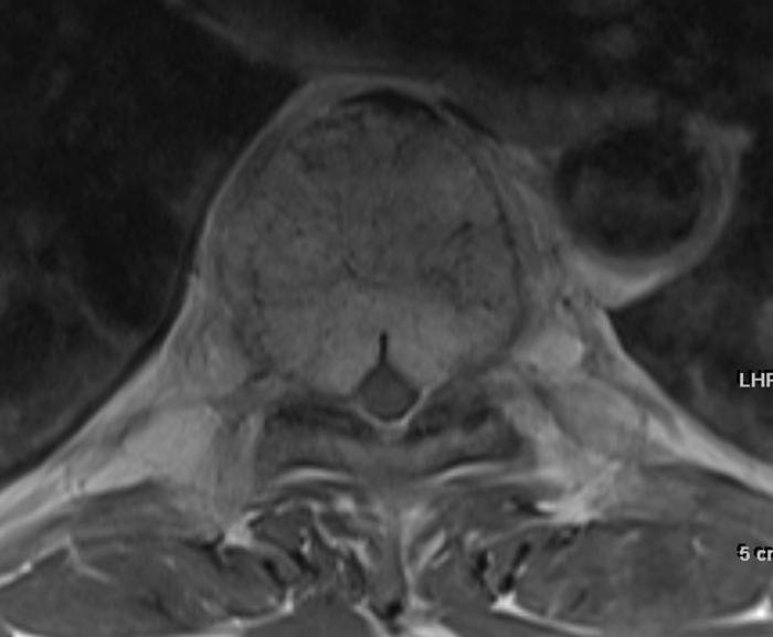
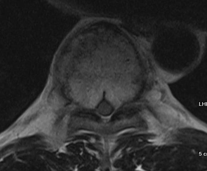
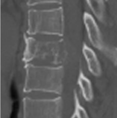
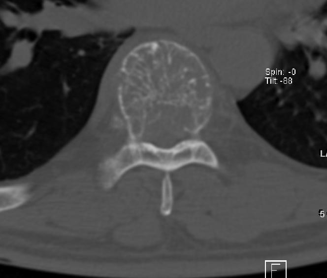
| Clinical information | 54/M, C.C : Both leg weakness on walking, hypoesthesia below T8 dermatome on walking for 1 month, |
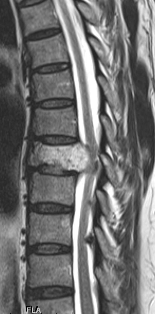
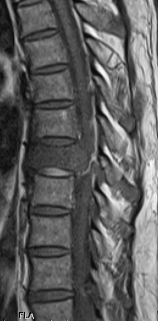
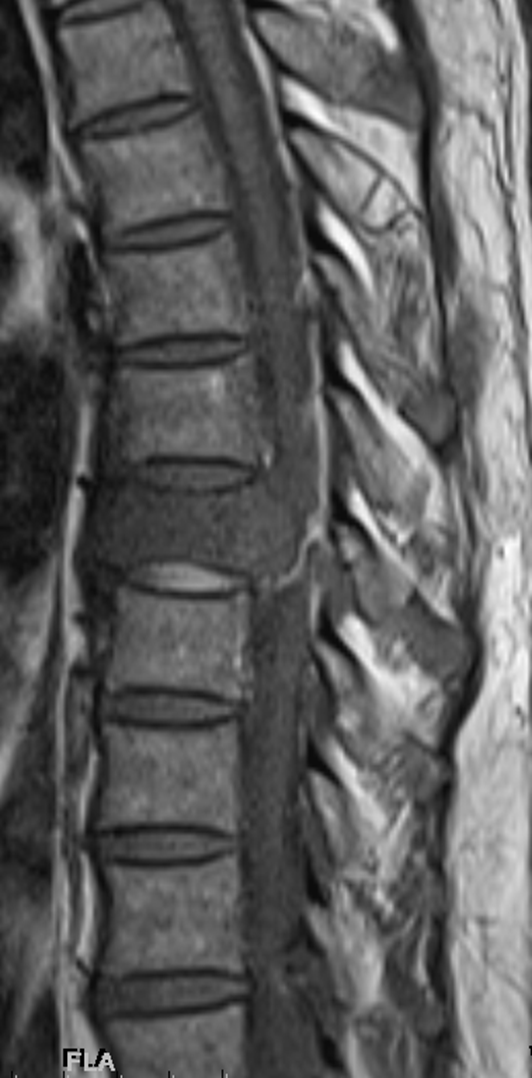
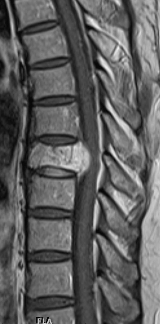
| Discussion | Aggressive hemangioma 사진 해설> T2 에서는 high T1 에서는 low signal intensity 를 보이는 lesion 이 thoracic vertebra body 를 replacement하고 있고 spinal canal 로 retropulsion 하고 있는 양상이다. 이 mass는 또한 strong 한 enhancement pattern을 보이고 있다. MRI finding 을 보았을 때는 plasmacytoma나 metastasis 와 같은 malignant lesion 을 생각할 수 있지만 Polka dot appearance의 CT finding 을고려하였을때 hemangioma를 생각하여야 하면 pathology 상 aggressive hemangioma가 확진 되었다. Review> The small but significant subset of symptomatic lesions are known as aggressive hemangiomas and they are characterized by bone expansion, extraosseous extension of tumor, disturbance of local blood flow and rarely compression fractures. Intralesional fat of hemangioma increased signal intensity on T1WI. On T2WI, the SI increases due to high water content. Aggressive hemangioma typically contain less fat and more vascular stroma thereby producing a low MR signal on T1WI, resembling metastatic lesion. Howevever, hemangioma is characterized by sparing and thickening of vertically striated trabeculae which preserve the functional capability of the vertebral body to withstand an axial load. This appearance has been described on CT as “ polka dot”, “honey comb” [1] References 1. Schrock WB, Wetzel RJ, Tanner SC, Khan MA (2011) Aggressive hemangioma of the thoracic spine. J Radiol Case Rep 5: 7-13. |
| Correct answer |
Correct Answer
Semi-Correct Answer |