HOME > Weekly Case > Weekly Case
Weekly Case
종결
Case No. 300
2013-04-01~2013-04-30
출제자 : 최승홍
Hit : 466
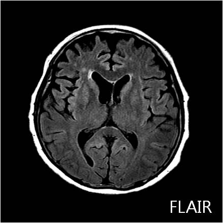
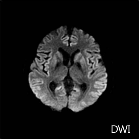
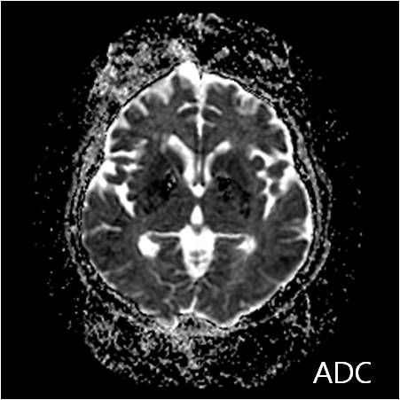
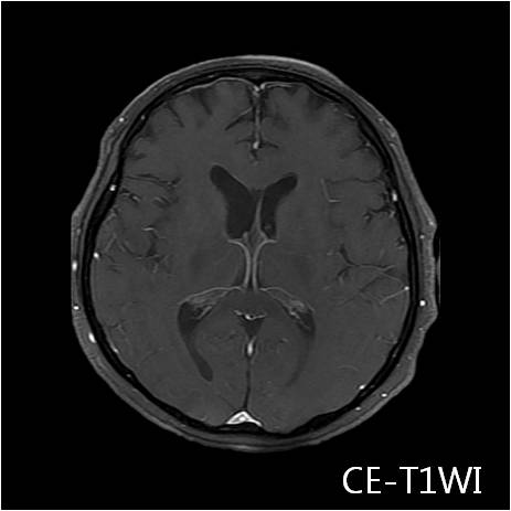
| Period | 2013-04-01~2013-04-30 |
|---|---|
| Diagnosis | CJD |
| Clinical information | F/65 CC: cognitive impairment (onset: 2 MA) 진단은? |
| Discussion | Terminology • CJD: Rapidly progressing, fatal, potentially transmissible dementia caused by a prion Imaging • Best imaging clue: Progressive T2 hyperintensity of basal ganglia (BG), thalamus, and cerebral cortex • Predominantly gray matter (GM): Caudate and putamen > GP o Thalamus: Common in variant CJD (vCJD) o Cerebral cortex: Frontal, parietal, and temporal • Heidenhain variant: Occipital lobe • 2 signs characteristic of vCJD o "Pulvinar" sign: Symmetric T2 hyperintensity of pulvinar of thalamus relative to anterior putamen o "Hockey stick" sign: Symmetric pulvinar and dorsomedial thalamic nuclear hyperintensity • Best imaging tool: MR with DWI Top Differential Diagnoses • Hypoxic-ischemic injury • Osmotic demyelination syndrome • Other causes of dementia o Alzheimer, frontotemporal, multi-infarct dementia, dementia in motor neuron disease • Leigh syndrome • Corticobasal degeneration Clinical Issues • Definite CJD diagnosed by brain biopsy or autopsy • Progressive dementia associated with myoclonic jerks & akinetic mutism; variable constellation of pyramidal, extrapyramidal, and cerebellar signs • Incidence 1 per 1,000,000 (USA and internationally) o sCJD (85%), familial (15%), infectious/iatrogenic (< 1%) (includes vCJD) • Death usually ensues within months of onset Reference: Diagnostic Imaging: Brain: 2nd edition by Anne Osborn, MD, FACR |
| Correct answer |
Correct Answer
Semi-Correct Answer |