HOME > Weekly Case > Weekly Case
Weekly Case
종결
Case No. 298
2013-03-04~2013-03-31
출제자 : 최현석
Hit : 528
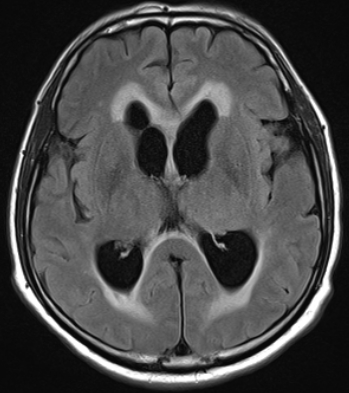
| Period | 2013-03-04~2013-03-31 |
|---|---|
| Diagnosis | Neurocysticercosis |
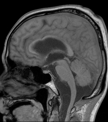
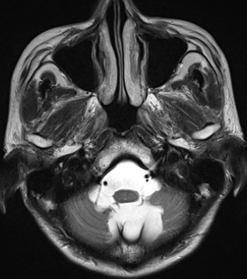
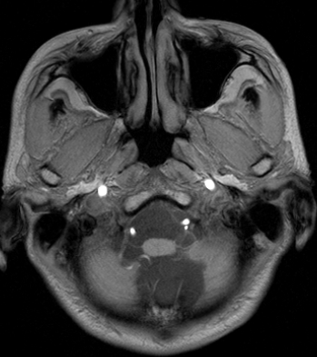
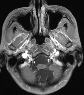
| Clinical information | 54/M Headache, Diplopia 진단은? |
| Discussion | Neurocysticercosis Cysticercosis (유구낭미충증) : m/c parasitic infection Location : CNS, eyes, muscles, subcutaneous tissue CNS lesion (60~90%) Neurocysticercosis Location - Subarachnoid-cisternal neurocysticercosis (m/c) - Parenchymal neurocysticercosis (2nd m/c) - Intraventricular neurocysticercosis Stage 1) Vesicular stage - A smooth thin-walled cyst isodense/isointense to CSF - Scolex : discrete, nodular, and hyperintense - No enhancement, no surrounding edema 2) Colloidal vesicular stage - Cyst fluid : hyperdense, hyperintense on T1, T2WIs - Scolex : hyperintense on FLAIR - Intense and “shaggy” enhancement of cyst wall - Surrounding edema 3) Granular nodular stage - Nodular or faint ring-like enhancement - Mild residual edema 4) Nodular calcified stage - Shrunken calcified nodule - No surrounding edema, no enhancement “Racemose” NCC - Multilobulated, variably sized, grape-like lesions in the basal cisterns - Lack an identifiable scolex - Arachnoiditis with fibrosis and scarring - rim enhancement around the cysts and along the brain surfaces - Obstructive hydrocephalus |
| Correct answer |
Correct Answer
Semi-Correct Answer |