HOME > Weekly Case > Weekly Case
Weekly Case
종결
Case No. 296
2013-03-04~2013-03-31
출제자 : 최현석
Hit : 370
| Period | 2013-03-04~2013-03-31 |
|---|---|
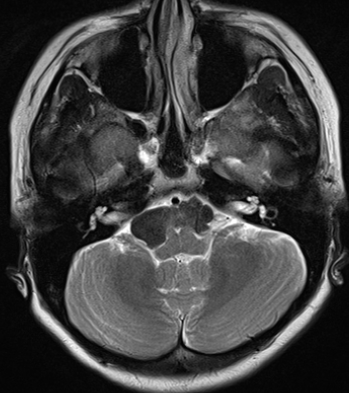
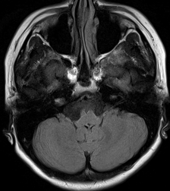
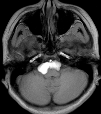
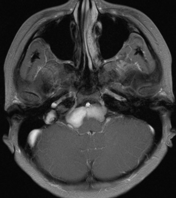
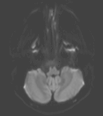
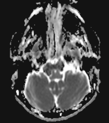
| Diagnosis | white epidermoid |
| Clinical information | Headache F/22 진단은? |
| Discussion | Intracranial epidermoid cyst Congenital inclusion cyst Intradural extraaxial location (>90%) - CPA cistern > middle cranial fossa / parasellar region > ventricles - Off-midline CT - Hypodense and identical to CSF - Calcification (10~25%) - Hemorrhage (very rare) MR - Iso- or slightly hyperintense to CSF on T1- and T2-Wis - No complete suppression on FLAIR, hyperintensity on DWI : DDx. Arachnoid cyst “white” epidermoid - High protein content - Hyperintense on T1WI, variable on T2WI - Hyperdense on CT DDx. 1) Arachnoid cyst - Isointense to CSF on all standard sequences - Completely nulls on FLAIR - No restricted diffusion - Displace local structures >> insinuate and engulf 2) Inflammatory cyst (neurocysticercosis) - Often enhances - Adjacent edema, gliosis 3) Cystic neoplasm - Attenuation/ SI not that of CSF, Enhancement (+) 4) Dermoid cyst - Usually midline - Fat (not CSF), Dermal appendage - Often ruptured |
| Correct answer |
Correct Answer
Semi-Correct Answer |