HOME > Weekly Case > Weekly Case
Weekly Case
종결
Case No. 294
2013-02-04~2013-02-28
출제자 : 최진욱
Hit : 442
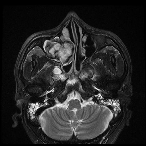
| Period | 2013-02-04~2013-02-28 |
|---|---|
| Diagnosis | Organizing hematoma of maxillary sinus |
| Clinical information | 33/M Past history : 두경부 종양으로 RT 받았다고 함. 진단은? |
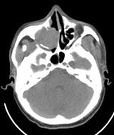
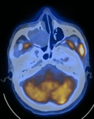
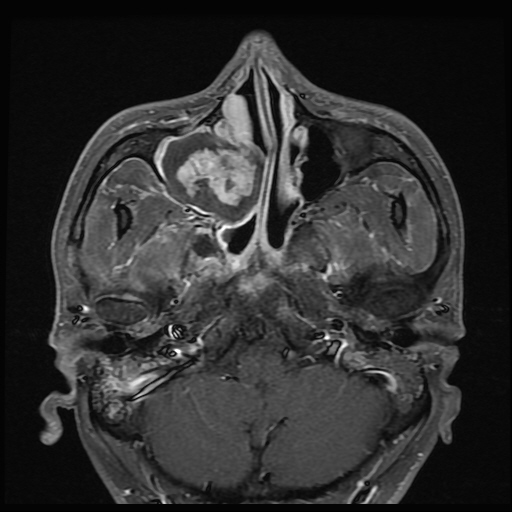
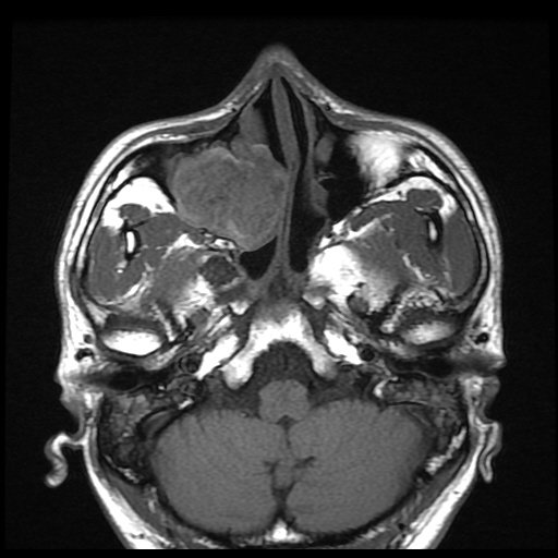
| Discussion | Organizing hematoma of maxillary sinus - Rare tumor like condition / Unilateral, medial antral wall near the sinus ostium - Underlying condition : bleeding diathesis, aggressive fungal infection, radiation therapy, a postop complication, or trauma - Pathogenesis : fibrosis and neovascularization -> repeated hemorrhage in the semiclosed lumen -> hematoma encapsulated by fibrosis -> prevents the absorption of the hematoma -> induces vascularization -> recurrent intra-capsular bleeding -> expansile mass formation - Clinical manifestation : repeated epistaxis(70%), congestion, rhinorrhea because of the obstruction and secondary inflammatory process, pain, facial swelling - Imaging findings of organizing hematoma Large well defined soft tissue mass causing marked expansion of the maxillary sinus with associated bone destruction CT imaging expansile maxillary sinus mass with some areas of bone resorption Heterogenous high densities and patch enhancement MR imaging variable signal intensity on T1- and T2WI Biphasic appearance – central T2 high, well enhancing, peripheral low SI DDX from malignancy Expansive but clearly demarcated, never infiltrative Compressive bony remodeling, but never destruction Well enhancing mucosal thickening suggesting inflammatory change, not destruction or invasion |
| Correct answer |
Correct Answer
Semi-Correct Answer |