HOME > Weekly Case > Weekly Case
Weekly Case
종결
Case No. 292
2013-01-01~2013-01-31
출제자 : 황승배
Hit : 459
| Period | 2013-01-01~2013-01-31 |
|---|---|
| Diagnosis | Spinal cord cavernous malformation |
| Clinical information | 16세 남자 C/C : Both lower extremity weakness and back pain (sudden onset) |
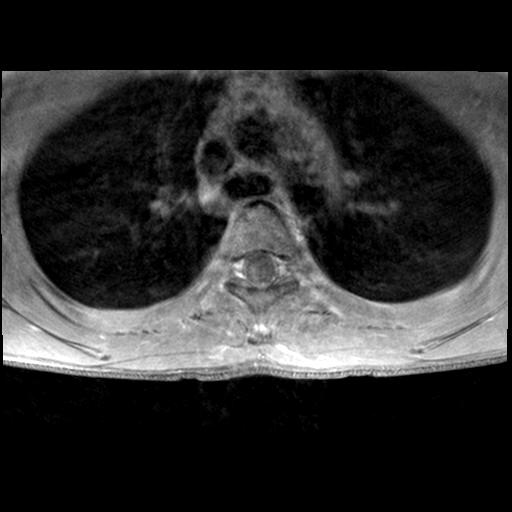
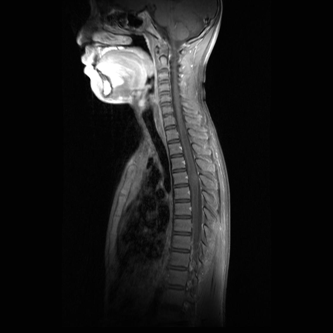
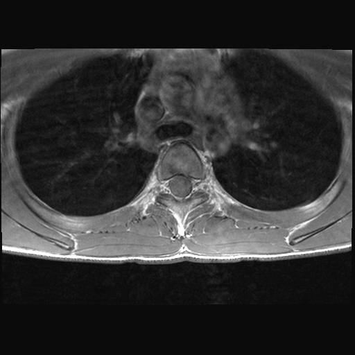
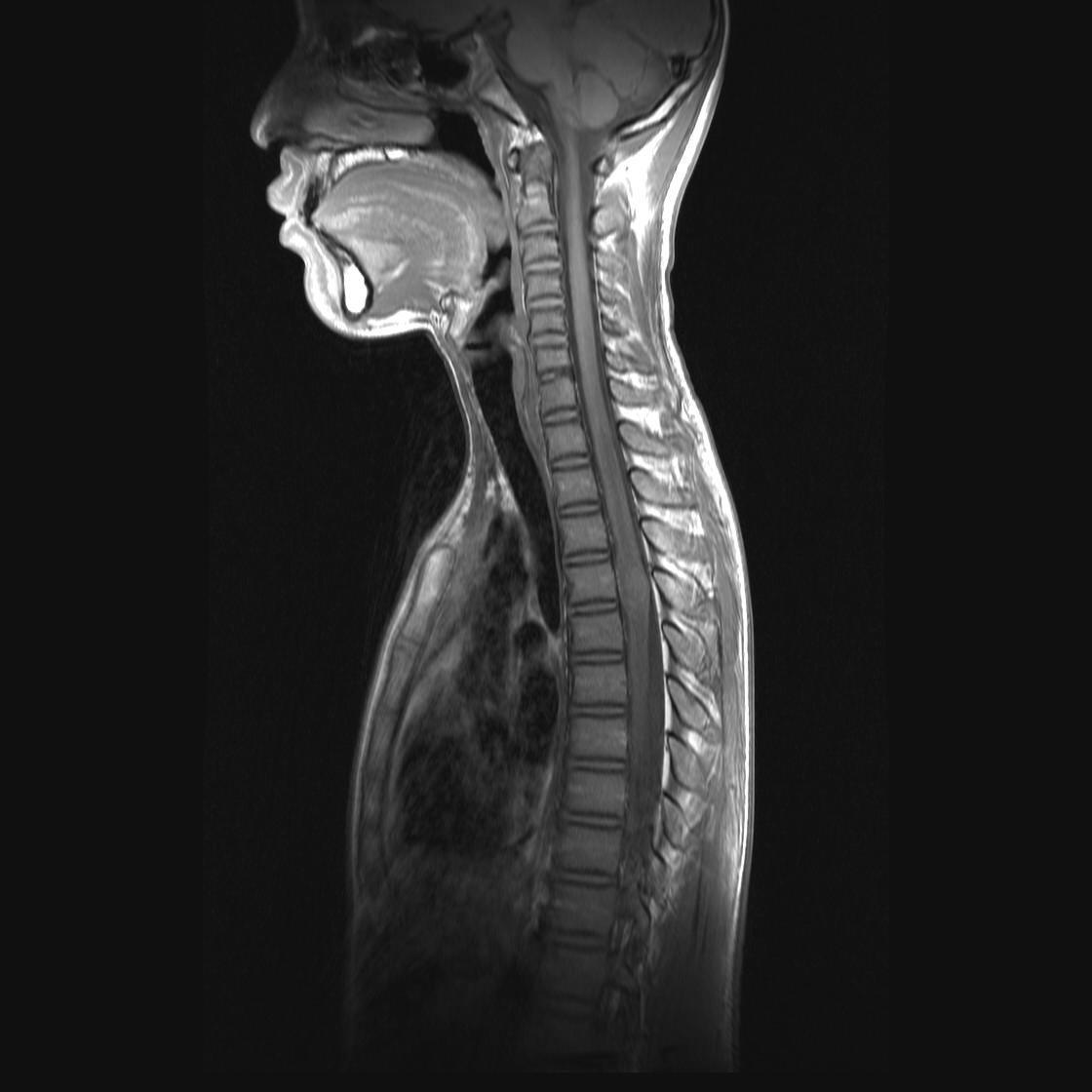
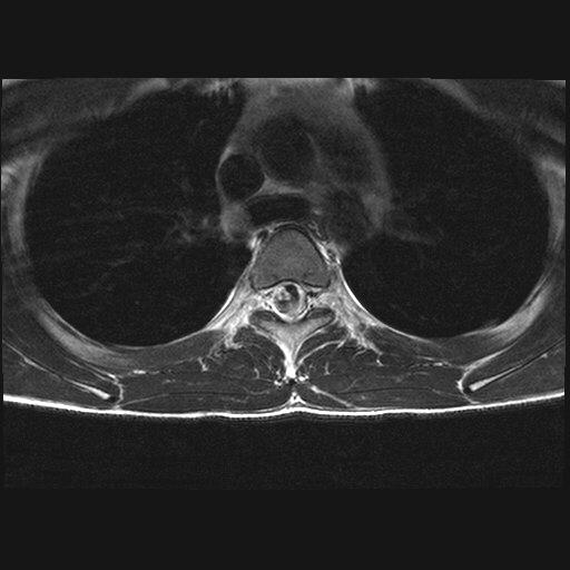
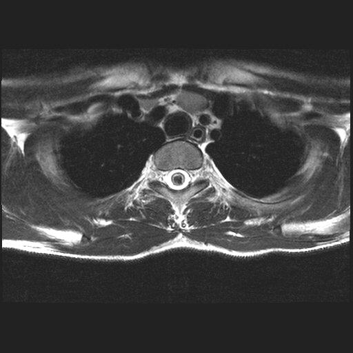
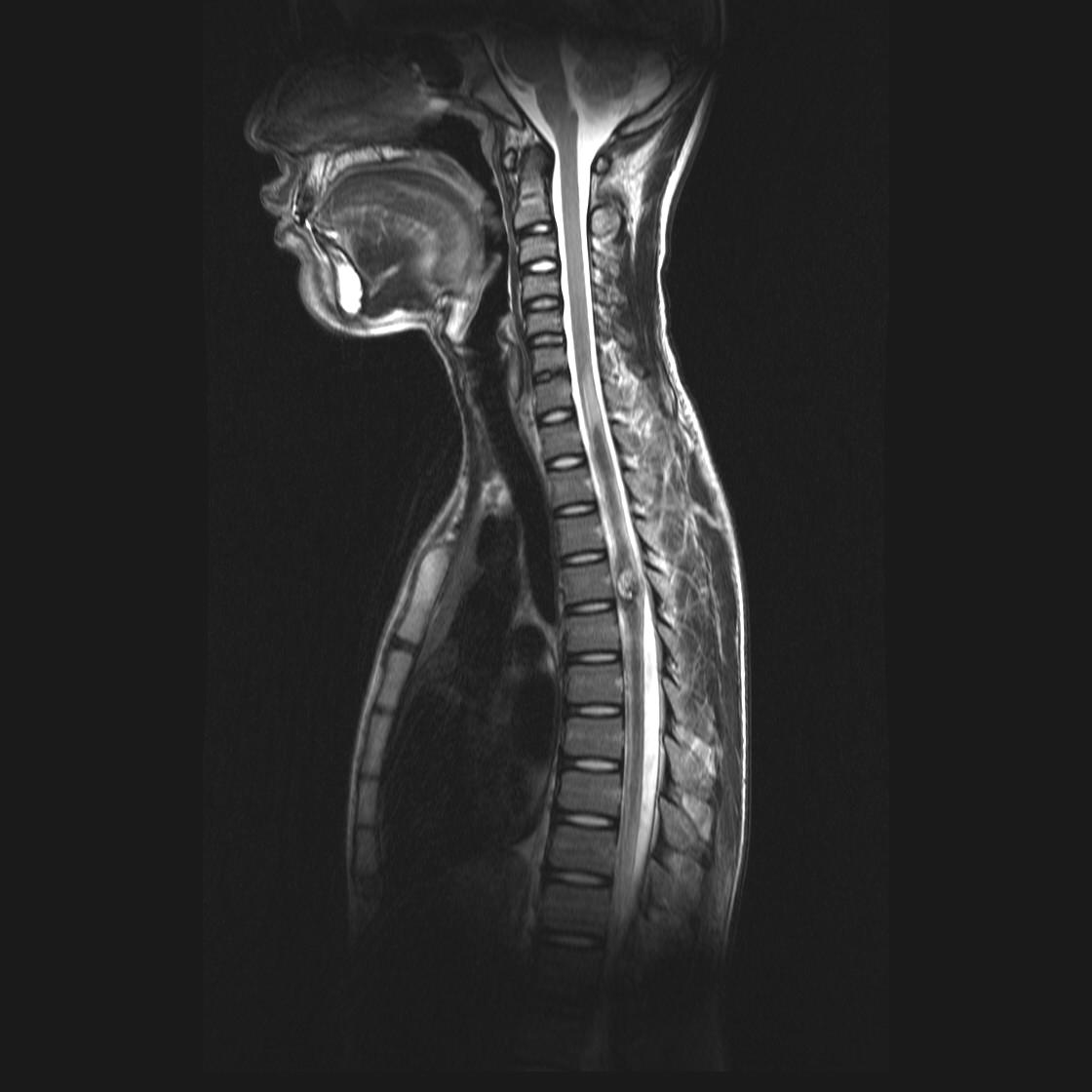
| Discussion | 사진설명 : T4 level의 spinal cord에 T2WI에서 mixed (high & low) signal intensity의 mass like lesion이 있으며 종괴주변으로 hemorrhage에 의한 dark signal intensity가 있고 가장 peripheral portion으로 edema에 의한 high signal intensity가 동반되어 있음. T1WI에서는 약간 heterogeneous isosignal intensity를 보이고 조영증강은 거의 관찰되지 않음. Spinal cord cavernous malformation Vascular lesion with lobulated, thin sinusoidal vascular channels, no interspersed neural tissue Locules of blood with fluid-fluid levels surrounded by very T2 hypointense rim Location ; spinal cord uncommon site, 3-5% of all cavernous malformation - Thoracic (50%) - Cervical (40%) - Conus (10%) Round heterogenous signal abnormality, well-defined margins T2WI - Heterogeneous (blood products, varying ages) - No edema, unless recent hemorrhage T1WI C+ ; enhancement absent/minimal Calcification ; rare (common in brain cavernous malformation) Bleeding rate ; 1-5% |
| Correct answer |
Correct Answer
Semi-Correct Answer |