HOME > Weekly Case > Weekly Case
Weekly Case
종결
Case No. 291
2013-01-01~2013-01-31
출제자 : 황승배
Hit : 381
| Period | 2013-01-01~2013-01-31 |
|---|---|
| Diagnosis | Achondroplasia |
| Clinical information | 1세 여아 C/C : Macrocephaly, developmental delay |
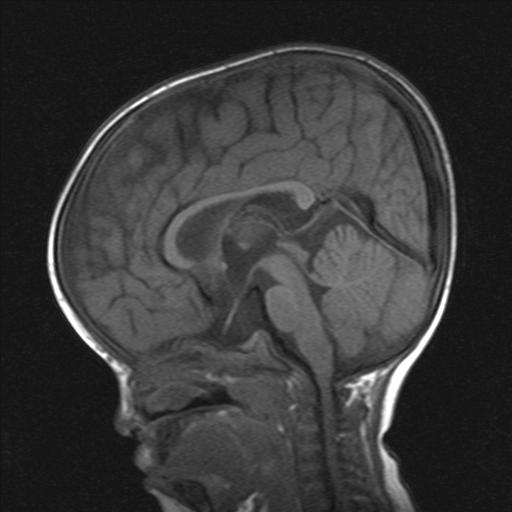
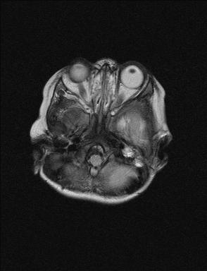
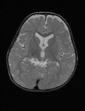
| Discussion | 사진설명 : Skull base가 형성이 덜 되어 있고 clivus도 작음. Foramen magnum이 좁고 cervicomedullary junction이 약간 compression되어 보임. Optic nerve가 stretching되어 있고 vertical course를 보임. Pituitary stalk이 stretching되어 잘 보이지 않음. Brainstem이 정상에 비해 upward displacement되어 있고 suprasellar sellar cistern이 넓어져 있음. Straight sinus가 정상에 비해 약간 커져 보임. Bifrontal subarachnoid space가 약간 넓어 보이며 mild ventricular enlargement가 있음. Achondroplasia Endochonral bone formation is inhibited The base of skull, the only part of the cranium formed by endochonral bone, is affected whereas the membranous bones of the calvaria develop and grow normally. => results in a relatively large skull with a small base and stenosis of the foramen magnum Cranial MR findings of achondroplasia Narrowing of the subarachnoid space at the level of foramen magnum Compressive deformities of the cervicomedullary junction ; foci of abnormal signal intensity in the upper cervical cord and medulla Upward displacement of the brainstem Enlargement of the suprasellar cistern Vertical course of the optic nerve Stretching of optic nerve and pituitary stalk Dilated lateral and third ventricles Bifrontal widening of the subarachnoid space Asymmetry in the outline of the cranium and brain Larger and more vertical course of the straight sinus Reference Kao SC, Waziri MH, Smith WL, et al. MR imaging of the craniovertebral junction, cranium, and brain in children with achondroplasia. AJR Am J Roentgenol 1989:153;565-569 |
| Correct answer |
Correct Answer
Semi-Correct Answer |