HOME > Weekly Case > Weekly Case
Weekly Case
종결
Case No. 282
2012-09-01~2012-09-30
출제자 : 최영준
Hit : 507
| Period | 2012-09-01~2012-09-30 |
|---|---|
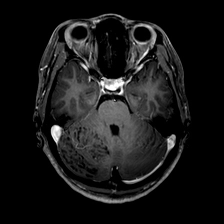
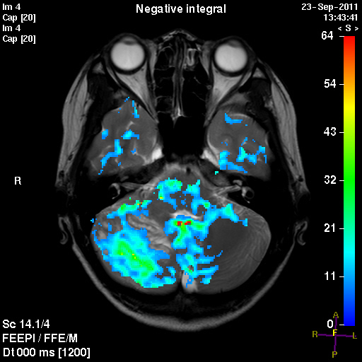
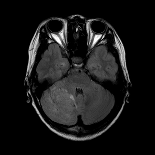
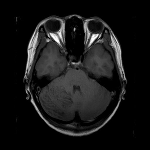
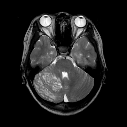
| Diagnosis | Lhermitte-Duclos Disease (Dysplastic Cerebellar Gangliocytoma) |
| Clinical information | F / 19, 주증상 : dizziness |
| Discussion | Lhermitte-Duclos Disease (Dysplastic Cerebellar Gangliocytoma) Lhermitte and Duclos disease(LDD) is a rare hamartomatous disorder. Lhermitte and Duclos reported the first case of cerebellar ganglion cell tumor in 1920, and this disease is now called dysplastic cerebellar gangliocytoma. Dysplastic cerebellar gangliocytoma is seen most frequently in young adults (average age, 34 years). Less frequently, it occurs in pediatric patients. There is no sex predilection. Patients present with headaches, occlusive hydrocephalus, cranial nerve palsies, gait ataxia and other symptoms of cerebellar dysfunction. Clinical evidence and the close association of dysplastic cerebellar gangliocytoma with Cowden syndrome, which is an autosomal-dominant phacomatosis and cancer syndrome, and this ‘multiple hamartoma-neoplasia syndrome’ is associated with mutations of the PTEN gene. MR imaging is the modality of choice. MR imaging reveals a cerebellar mass with a typical striated or tiger-striped folial pattern that consists of alternating bands on both T1- and T2-weighted images. The bands are hyper- and isointense relative to gray matter on T2-weighted images and iso- and hypointense on T1-weighted images. Most dysplastic gangliocytomas do not enhance; however, enhancement has been reported and is probably due to the presence of anomalous veins. Mass effect is common and causes compression of the fourth ventricle and oc- clusive hydrocephalus. Reference : Shinagare AB, Patil NK, Sorte SZ. Case 144: Dysplastic cerebellar gangliocytoma (Lhermitte-Duclos disease). Radiology 2009; 251:298–303 Moenninghoff C, Kraff O, Schlamann M, Ladd ME, Katsarava Z, Gizewski ER. Assessing a Dysplastic Cerebellar Gangliocytoma (Lhermitte-Duclos Disease) with 7T MR Imaging, Korean J Radiol 2010;11:244-248 |
| Correct answer |
Correct Answer
Semi-Correct Answer |