HOME > Weekly Case > Weekly Case
Weekly Case
종결
Case No. 280
2012-08-01~2012-08-31
출제자 : 최진우
Hit : 497
| Period | 2012-08-01~2012-08-31 |
|---|---|
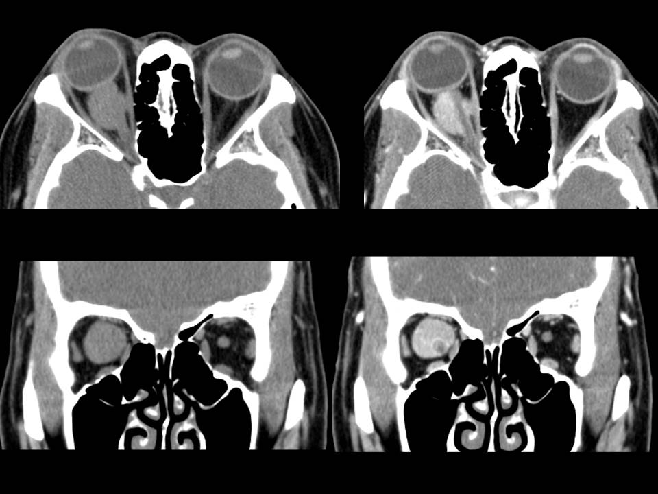
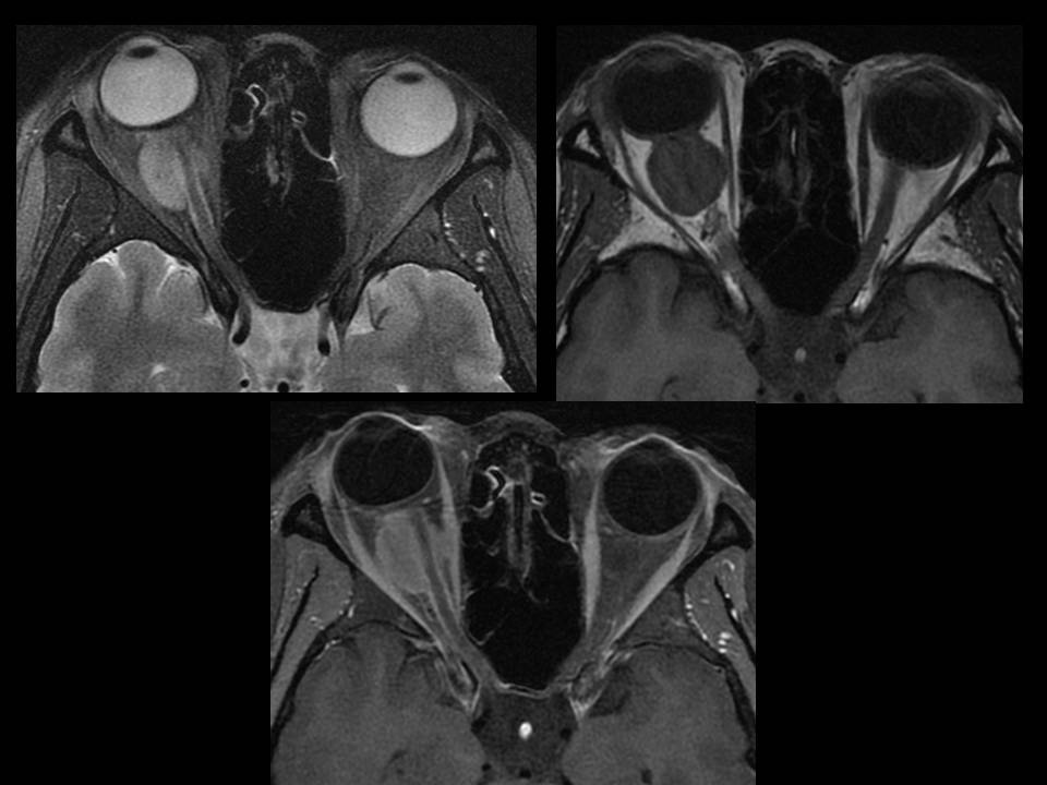
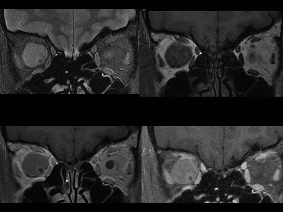
| Diagnosis | Optic nerve sheath meningioma, transitional type |
| Clinical information | 46/F C/C : proptosis, right onset) 1 MA |
| Discussion | Dx: Optic nerve sheath meningioma, transitional type, WHO grade I/IV REVIEW: Optic nerve sheath meningioma 1. rare tumors of the anterior visual pathway, 2% of all orbital tumors 2. Bilateral and multifocal presentations of ONSMs are most commonly found in patients with neurofibromatosis type 2. 3. Arising from arachnoid cap cells within the optic nerve sheath 4. Age: fourth and fifth decades of life 5. In pediatric patient : more aggressive, high rate of intracranial extension and recurrence, bilateral presentation 6. Sx: painless, progressive loss of visual acuity or visual field, proptosis Imaging feature Tubular(m/c), globular, fusiform, and focal pattern CT 1. Homogeneous and a smooth or slightly lobulated peripheral margin 2. isodense to the optic nerve MR 1. isointense to the optic nerve on T1- and T2WI 2. Homogeneous enhancement (comparable to EOM) 3. “Tram track” sign : a relatively lucent optic nerve in the center of an enlarged ONS complex with peripheral enhancement References 1. Optic nerve sheath meningioma: current diagnosis and treatment. Neurosurg Focus 23 (5):E4, 2007 2. Radiologic-Pathologic Correlation Meningioma of the Optic Nerve Sheath. AJNR 17:901–906 |
| Correct answer |
Correct Answer
Semi-Correct Answer |