HOME > Weekly Case > Weekly Case
Weekly Case
종결
Case No. 278
2012-08-01~2012-08-31
출제자 : 최진우
Hit : 455
| Period | 2012-08-01~2012-08-31 |
|---|---|
| Diagnosis | Dysembryoplastic neuroepithelial tumor |
| Clinical information | 12/F C/C : Seizure onset) 2~3 MA |
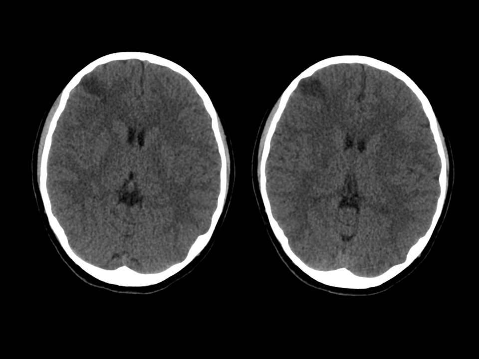
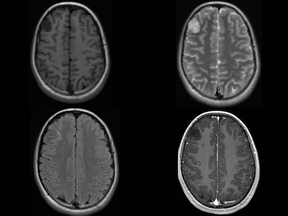
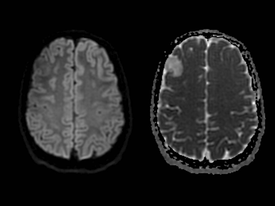
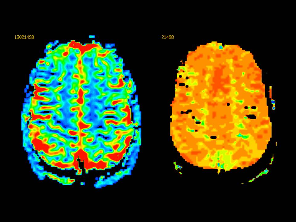
| Discussion | Dx: Dysembryoplastic neuroepithelial tumor, WHO grade I DNT 1. A benign, usually supratentorial, mixed neuronal-glial neoplasm 2. Multinodular architecture and a predominantly intracortical location 3. Composed of heterogeneous cellular components, such as oligodendrocyte like cells with admixtures of mature ganglion cells and astrocytes, which are located in myxoid or mucinous matrix 4. Frequently associated with cortical dysplasia Radiologic Findings CT 1. Moderately or markedly hypoattenuating, wedge-shaped cyst like lesion 2. Scalloped inner table in 44-60%, calcification in less than 20% MRI 1. Well-demarcated, multilobulated(“bubbly”) or gyriform appearance 2. Hypointense on T1WI, markedly hyperintense on T2WI 3. Well defined, complete or incomplete hyperintense ring on FLAIR 4. High frequency of bone remodeling of the adjacent calvaria 5. No surrounding vasogenic edema and enhancement References Neuronal Tumors of the Central Nervous System: Radiologic Findings and Pathologic Correlation : RadioGraphics 2002; 22:1177–1189 Diagnostic neuroradiology : V. N. Kornienko • I. N. Pronin , 2009 Springer |
| Correct answer |
Correct Answer
Semi-Correct Answer |