HOME > Weekly Case > Weekly Case
Weekly Case
종결
Case No. 273
2012-06-01~2012-06-30
출제자 : 김진아
Hit : 438
| Period | 2012-06-01~2012-06-30 |
|---|---|
| Diagnosis | Heterotopia |
| Clinical information | 26세 남자, seizure for 7 years. 진단은? |
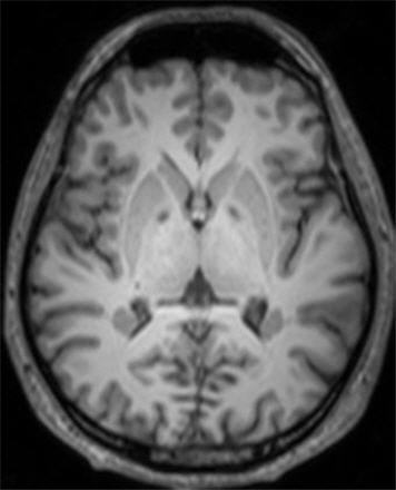
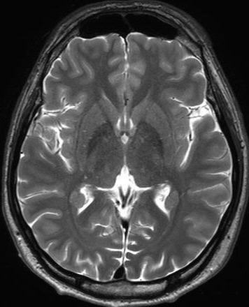
| Discussion | Heterotopia Heterotopia represents collections of normal neurons located in abnormal locations, anywhere from the subependymal region to the cerebral cortex. They result from the arrest of neuroblast migration. Patients commonly present with epilepsy and possible delayed milestones and/or mental retardation. Heterotopia within the brain is often divided into three groups: subependymal heterotopia, focal cortical heterotopia, and band heterotopia Periventricular (Subependymal) Heterotopia: Periventricular (subependymal) heterotopias (PVH) are located in close proximity to the ventricular wall. They are commonly seen in the region of the trigone and occipital horns of the lateral ventricles. PVH are usually bilateral with predilection for the right cerebral hemisphere due to later migration of the right-sided neuroblasts. They appear as round or oval nodules, are isointense to the normal gray matter on all pulse sequences, and do not enhance after contrast injection. They may lie in the wall of the ventricle and project into the ventricular lumen or lie within the periventricular white matter. Mild ventricular dilation might be seen. Subcortical Heterotopia: Subcortical heterotopias (SCH) are located within the subcortical or deep white matter and are always seen contiguous to the overlying cortex or the underlying ventricular system. The affected hemisphere may decrease in size as a result of the decreased volume of the neurons. The overlying cortex appears thin with shallow sulci. SCH are either nodular, curvilinear, or mixed form. Band (Laminar) Heterotopia: Band heterotopia is a rare anomaly seen predominantly in females and may be familial, with X-linked dominant inheritance. It results from an early arrest of neuronal migration and consists of a smooth layer of the gray matter that often follows the curvature of the overlying cortex. On MR imaging, it shows the characteristic 3-layer cake (continuous double cortex) with the cortex and bilateral symmetric circumferential subcortical layer of band heterotopia separated from each other by a thin white matter band. The cortex may be relatively normal or pachygyric. References 1. Barkovich AJ, Raybaud CA. Neuroimaging in disorders of cortical development. Neuroimaging Clin N Am 2004;14:231–254,vii 2. Abdel Razek AA, Kandell AY, Elsorogy LG, Elmongy A, Basett AA. Disorders of cortical formation: MR imaging features. AJNR 2009;30:4-11 |
| Correct answer |
Correct Answer
Semi-Correct Answer |