HOME > Weekly Case > Weekly Case
Weekly Case
종결
Case No. 267
2012-04-02~2012-04-30
출제자 : 최승홍
Hit : 522
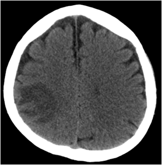
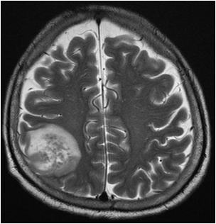
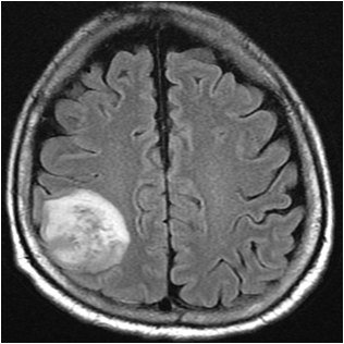
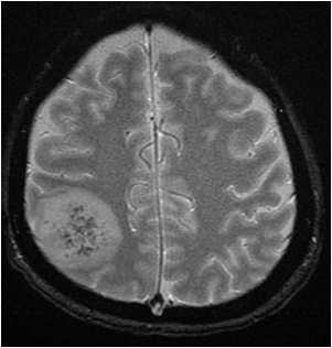
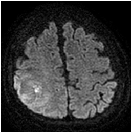
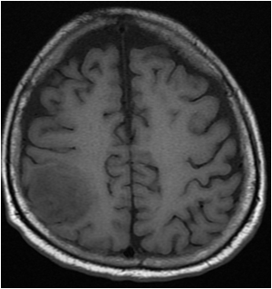
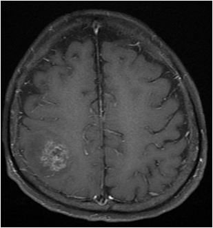
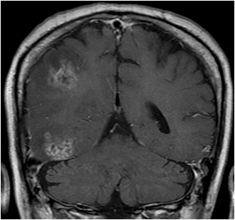
| Period | 2012-04-02~2012-04-30 |
|---|---|
| Diagnosis | Fungal (aspergiollosis) cerebritis |
| Clinical information | M/47 Hx: alcoholic LC, s/p liver TPL (1 week ago) CC: Sudden onset Lt. side weakness, Drowsy mentality |
| Discussion | Fungal (aspergiollosis) cerebritis Invasive aspergillosis of the central nervous system is becoming more common, particularly because of the increasing prevalence of immunosuppression and the increasing length of survival of immunosuppressed patients. Candida andAspergillus are among the most common fungal pathogens and are responsible for the majority of brain abscesses in immunocompromised patients. A Candida albicans brain abscess is usually caused by venous catheter–related candidemia.Aspergillus enters the body through the respiratory system by means of the inhalation of spores, most commonly those of Aspergillus fumigatus. They may then enter the central nervous system either hematogenously or directly from the paranasal sinuses. Another common opportunistic fungus, Cryptococcus neoformans, usually manifests with meningoencephalitis; it rarely forms a focal mass lesion in the brain parenchyma. Aspergillus abscesses generally appear hypointense on unenhanced T1-weighted MR images, but areas of high signal intensity may be seen. Shortened T1 relaxation times may be due to the presence of iron, manganese, or methemoglobin. Hemorrhage occurs in approximately 25% of lesions. Contrast enhancement is usually minimal or absent. Enhancement is thought to depend on the amount of inflammatory reaction, which varies with the stage of the infection and the immune status of the patient. On T2-weighted images, the areas of low signal intensity at the periphery of the lesions correspond, at least partially, to hemorrhage. This can best be seen on gradient-echo T2-weighted images. In a study of fungal sinus infections by Zinreich et al, the zones of low signal intensity on T2-weighted images were attributed to the presence of iron, manganese, and magnesium in the fungal concretions. Aspergillus abscesses have been shown to contain a dense population of hyphal elements peripherally, with a relative paucity of fungal elements centrally, features that likely contribute to the distinct peripheral low signal intensity on T2-weighted images. |
| Correct answer |
Correct Answer
Semi-Correct Answer |