HOME > Weekly Case > Weekly Case
Weekly Case
종결
Case No. 263
2012-03-02~2012-03-30
출제자 : 정원상
Hit : 443
| Period | 2012-03-02~2012-03-30 |
|---|---|
| Diagnosis | Nodular fasciitis |
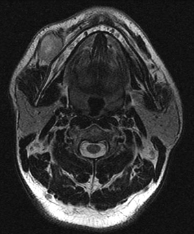
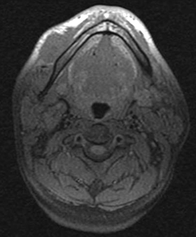
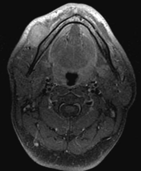
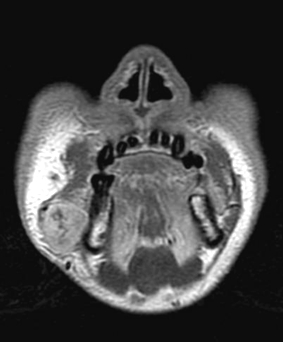
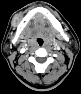
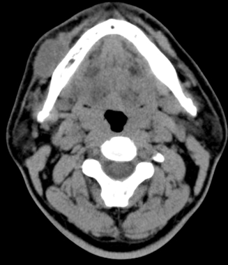
| Clinical information | 42/M C/C : 5개월 전부터 만져지는 right neck mass 점점 커지는 양상 진단은? |
| Discussion | Nodular fasciitis Benign tumor-like fibroblastic proliferation Subcutaneous pseudosarcomatous fibromatosis, infiltrative fasciitis, or proliferative fasciitis Nodular fasciitis can be found anywhere in the body : upper extremity (48%), trunk (20%), head and neck (15%–20%), and lower extremities (15%) Most common clinical presentation - solitary, rapidly growing mass with frequently associated pain and tenderness - trauma history CT and MR finding - Relatively well-defined, soft-tissue mass of superficial location - Deep-seated lesion인 경우 intramuscular type ; tend to be large and have ill-defined margins aggressive clinical behavior simulating a malignancy - various degrees of enhancement - various signal intensities in MR ; variability in cellularity, amount of collagen, amount of cytoplasm and water content Reference Kim ST, Kim HJ, Park SW et al. Nodular Fasciitis in the Head and Neck: CT and MR Imaging Findings. AJNR 2005;26:2617-2623 |
| Correct answer |
Correct AnswerSemi-Correct Answer |