HOME > Weekly Case > Weekly Case
Weekly Case
종결
Case No. 255
2011-10-04~2011-10-31
출제자 : 임현경
Hit : 548
| Period | 2011-10-04~2011-10-31 |
|---|---|
| Diagnosis | |
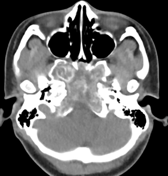
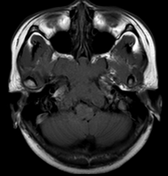
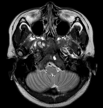
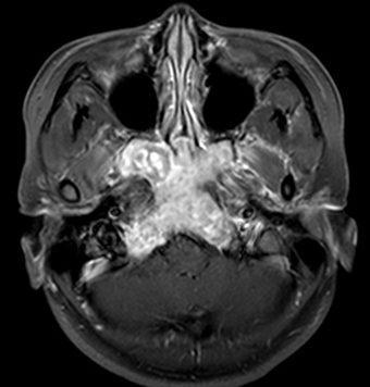
| Clinical information | 22/F Incidental lesion at the skull base 진단은? |
| Discussion | Fibrous dysplasia - Idiopathic skeletal developmental anomaly that can affect bone (monostotic form) or multiple bones (polyostotic form) - Often occurs early in life, with an average age of 10 years at onset : The lesions often stop enlarging after puberty - The skull base is commonly involved : Lesions of the skull can grow quite large and cause facial deformity, exophthalmos, and narrowing of skull base foramina - Radiologic finding o. plain films : classic ground-glass appearance o. CT: useful for skull base lesions, the characteristic ground-glass appearance, often with an associated sclerotic rim o. MR : Correlation with CT is critical to make the appropriate diagnosis homogeneously low signal intensity on T1WI and appear variable on T2WI o. Radionuclide bone scanning : may be useful for detecting early polyostotic disease and follow-up increased radionuclide uptake in both perfusion and static bone imaging - Prognosis Generally have a good prognosis Rarely, they may undergo spontaneous malignant transformation, and radiation therapy would increase this risk Reference 1. Y.W. Lui et al., Sphenoid Masses in Children: Radiologic Differential Diagnosis with Pathologic Correlation; American Journal of Neuroradiology 32:617-626, April 2011 2. Greenspan A, ed. Orthopedic Imaging: A Practical Approach. 4th ed. Philadelphia:Lippincott Williams & Wilkins; 2004 |
| Correct answer |
Correct Answer
Semi-Correct Answer |