HOME > Weekly Case > Weekly Case
Weekly Case
종결
Case No. 254
2011-10-04~2011-10-31
출제자 : 임현경
Hit : 520

| Period | 2011-10-04~2011-10-31 |
|---|---|
| Diagnosis | |
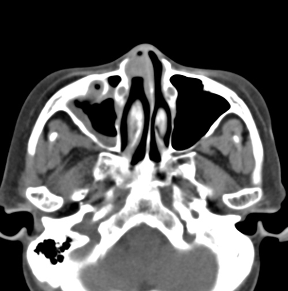
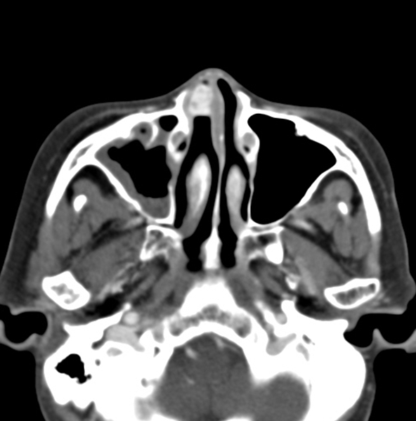
| Clinical information | 67/F C.C. : epistaxis 진단은? |
| Discussion | Pyogenic granuloma (lobular capillary hemangioma) of the nasal cavity Benign neoplasm of unknown etiology No recurrence Occurrence: unknown, all age but commonly in women of the third to fifth decades C.C.: unilateral epistaxis (95%), nasal obstruction (35%) Location: anterior nasal septum, inferior turbinate Size: 1-8cm (average 1.7cm) Pathology (2 layer) - Lobular area: capillary proliferation with microscopically distinct lobular architecture - Superficial ulcerative area: stromal edema, capillary dilatation, inflammation, granulation tissue reaction CT findings: - Lobular intense enhancing mass with iso- or hypoattenuating cap of variable thickness around intensely enhancing mass - Bone change: erosion (50%), displacement (33%) MRI findings - lowSI on T1WI and high SI on T2WI and intense enhancement D/Dx - juvenile angiofibroma, angiomatous polyp,hemangioma, hemangiopericytoma, paraganglioma, angiosarcoma, and hypervascularmetastases 3. reference -Puxeddu R, BerlucchiM, LeddaGP, et al. Lobular capillary hemangioma of thenasal cavity: a retrospective study on 40 patients. AmJ Rhinol 2006;20:480–484 - Lee DG, Lee SK, Chang HW, et al. CT features of locapillaryhemangioma of the nasal cavity. AJNR 2010;31:749-754 |
| Correct answer |
Correct Answer
Semi-Correct Answer |