HOME > Weekly Case > Weekly Case
Weekly Case
종결
Case No. 249
2011-08-01~2011-08-31
출제자 : 황승배
Hit : 591
| Period | 2011-08-01~2011-08-31 |
|---|---|
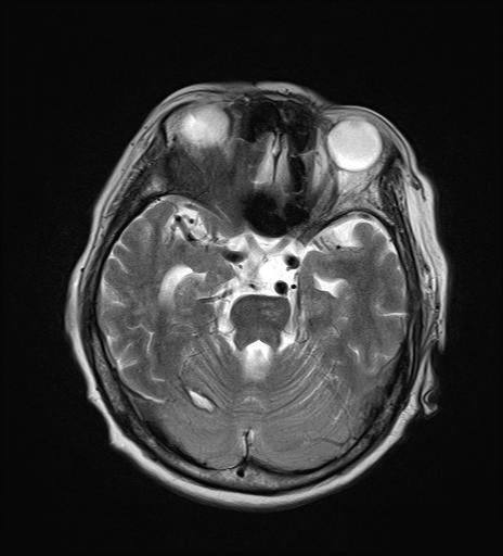
| Diagnosis | Remote cerebellar hemmorrhage |
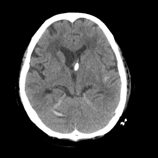
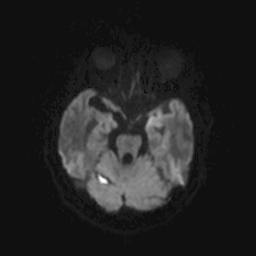
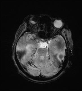
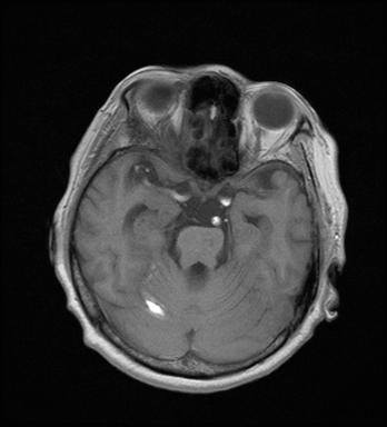
| Clinical information | 64세 여자 C/C : Rt side weakness & aphasia로 시행한 brain CT상 Lt thalamus에 acute ICH & IVH로 external drainage시행후 follow up brain CT 사진상 이상 소견 발견. MRI는 FU brain CT후 1주일후에 촬영 진단은? |
| Discussion | Remote cerebellar hemmorrhage * Complication of neurosurgical procedures - Most often after fronto-temporal or frontal craniotomies - Aneurysm clipping - Temporal lobectomy - Tumor resection - Hematoma evacuation * Pathophysiology 1) Supratentorial procedures that involve opening of cisterns or ventricular systems => CSF hypovolemia => Cerebellar sagging => Transient occlusion of the superior bridging veins of the posterior fossa => Hemorrhagic infarction 2) Negative suctioning => Postoperative over-drainage => Downward displacement of the cerebellum => Tearing of the superior cerebellar vein & tributaries * Can occur contralateral or ipsilateral to the site of craniotomy * Self-limiting phenomenon that should not be mistaken for more ominous pathologic findings * Imaging features “Zebra sign” : Locations - Sublocation in cerebellar vermis - Subarachnoid +/- superficial parenchymal bleed Contralateral to side of surgery (29%) Ipsilateral (22%) Bilateral (33%) Isolated vermian (9%) 참고문헌 1) AJNR Am J Neuroradiol 27:387–390 (2006) |
| Correct answer |
Correct AnswerSemi-Correct Answer |