HOME > Weekly Case > Weekly Case
Weekly Case
종결
Case No. 247
2011-07-04~2011-07-31
출제자 : 김진아
Hit : 507
| Period | 2011-07-04~2011-07-31 |
|---|---|
| Diagnosis | |
| Clinical information | 69세 여자, memory impairment 진단은? |
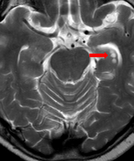
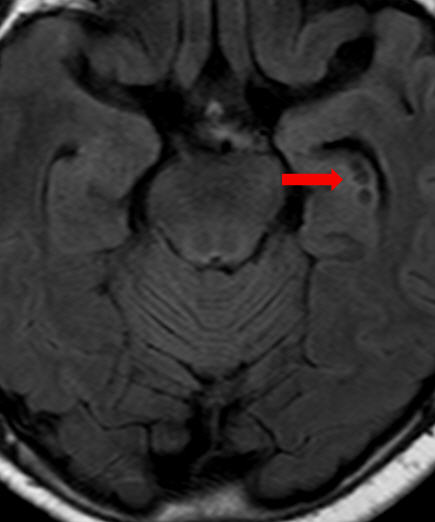
| Discussion | Hippocampal sulcus remnants Hippocampal sulcus remnants, which are also called hippocampal remnant cysts, are string of cysts along lateral margin of hippocampus between the cornu ammonis and the dentate gyrus. Primitive hippocampal fissure is formed as cornu ammonis and dentate gyrus fold on each other to form cleft. Cornu ammonis and dentate gyrus then fuse, leaving shallow hippocampal sulcus. Defects in this fusion cause remnant cysts within primitive sulcus. These are normal variants that are found in about 10-15% of seizure patients and have a higher incidence in older patients. The hippocampal sulcus remnants follow CSF signal on all MR sequences; hyperintense on T2WI, complete suppression on FLAIR, no diffusion restriction on DWI. They have been noted to enlarge with temporal lobe atrophy. Reference Sasaki M, Sone M, Ehara S, Tamakawa Y. Hippocampal sulcus remnant: potential cause of change in signal intensity in the hippocampus. Radiology 1993;188:743-6. |
| Correct answer |
Correct AnswerSemi-Correct Answer |