HOME > Weekly Case > Weekly Case
Weekly Case
종결
Case No. 233
2010-12-29~2011-01-13
출제자 : 관리자
Hit : 555
| Period | 2010-12-29~2011-01-13 |
|---|---|
| Diagnosis | |
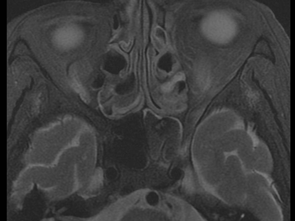
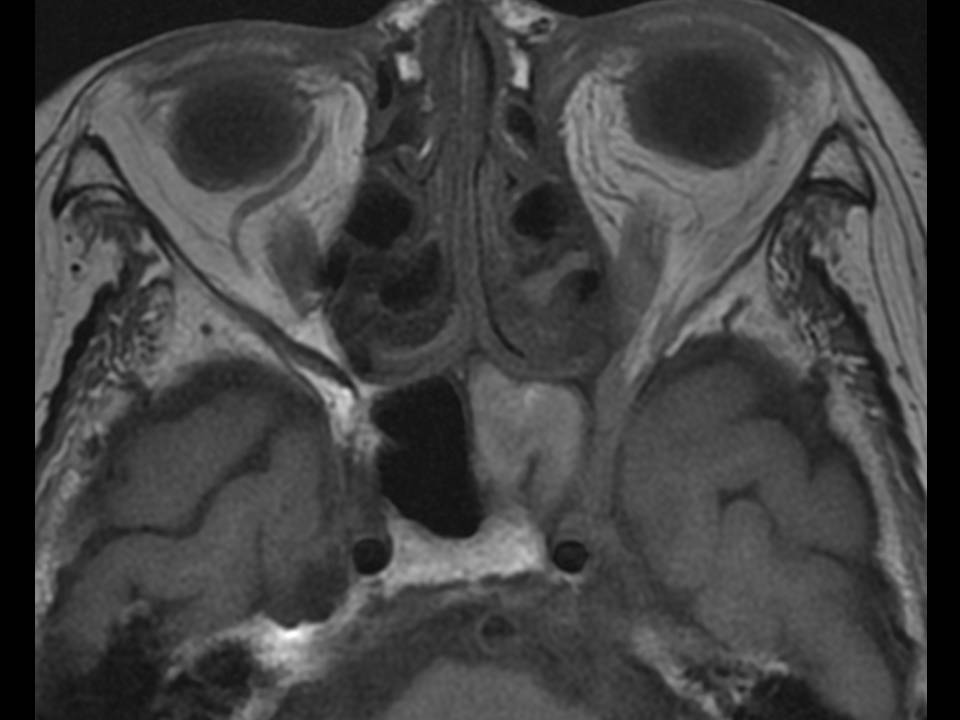
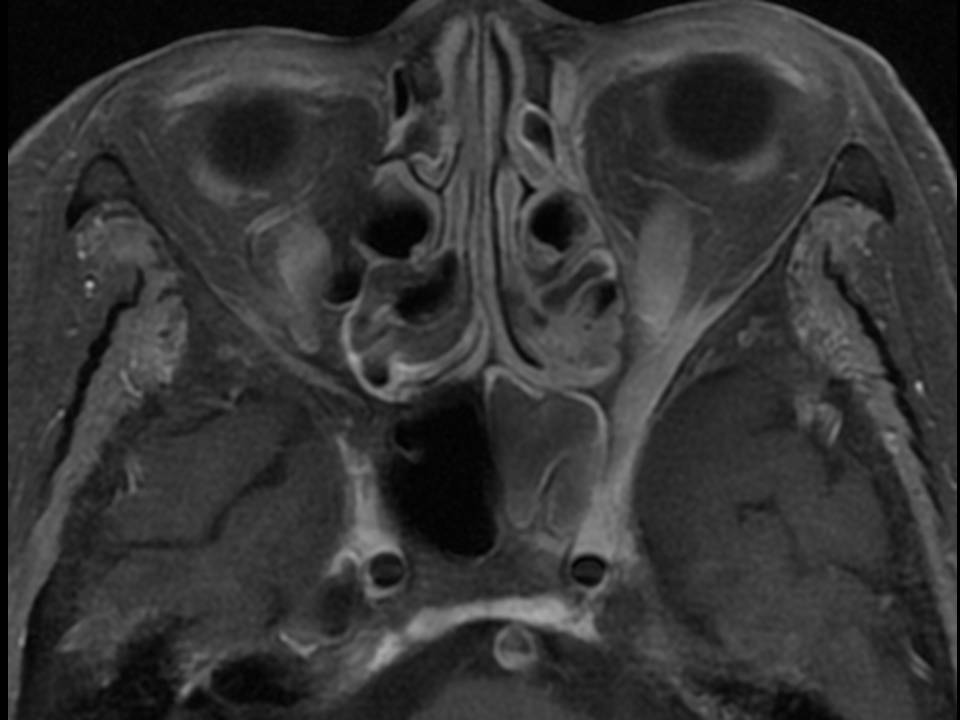
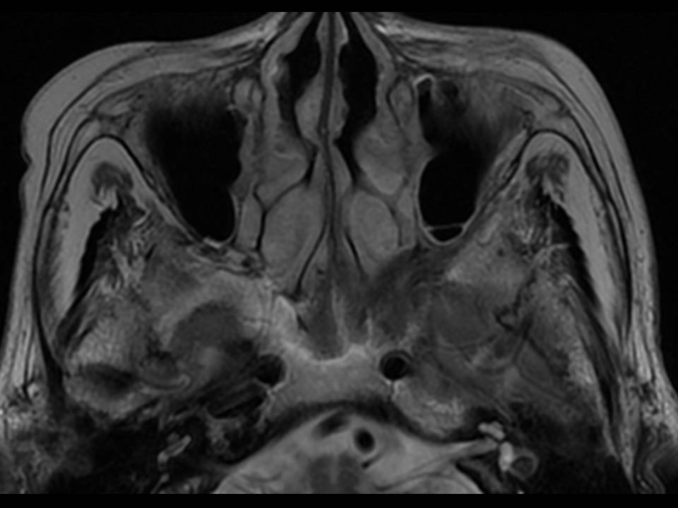
| Clinical information | F/88 C.C.: Diplopia PMHx: DM |
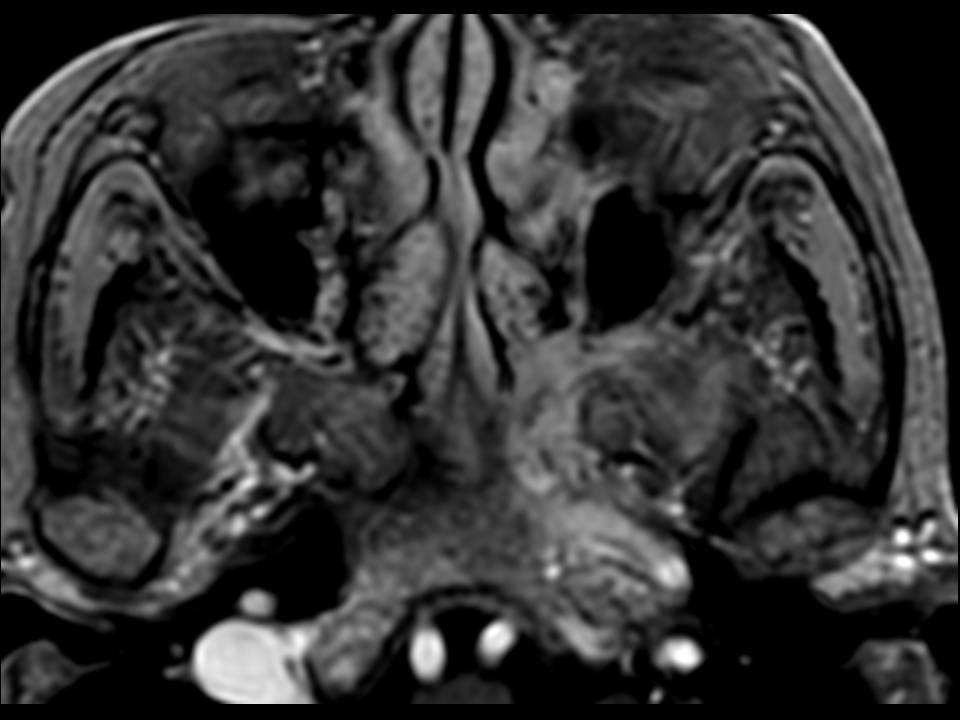
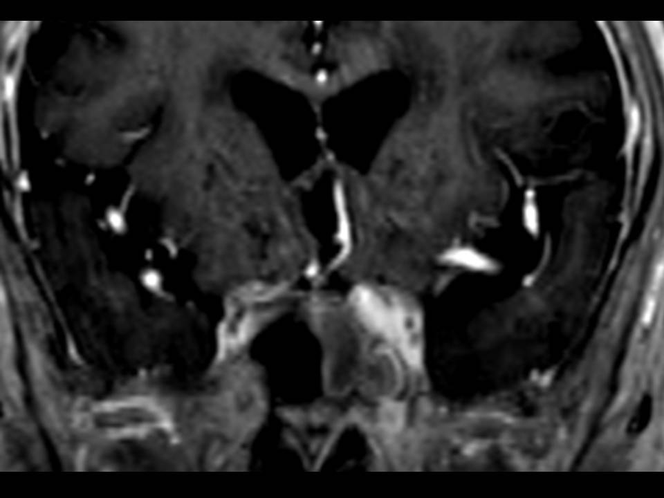
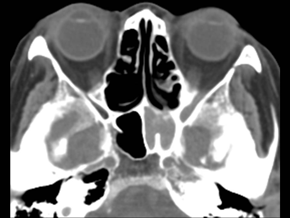
| Discussion | 정답: Invasive fungal sinusitis (Aspergillosis) 설명: Axial T2, T1WI에서 left sphenoid sinus에 T2 low, T1 iso to high signal intensity를 보이는 lesion이 가득 차 있으며 enhancement시 sphenoid wall을 따라서 enhancement를 보임. 동반하여 left orbital apex, pterygomaxillary fissure, inferior orbital fissure와 인접한 sphenoid bone을 따라서 T2WI에서 low signal intensity를 보이고 enhancement가 되는 infiltrative nature의 lesion들이 있음. Left cavernous sinus도 enhancement가 되고 bulging을 보임. CT에서 left sphenoid bone이 thickening되어 있으며 left sphenoid sinus내에 soft tissue density가 보이는데 center에 subtle한 high density area들도 보임. 고찰: A variety of fungal diseases such as aspergillosis, mucrormycosis, candidiasis, hitoplasmosis, cryptococcosis, coccidioidomycosis involve the sinonasal cavities. Insidious progression occurs over several months to years in which fungal organisms invade the mucosa, submucosa, blood vessels, and bony walls of the paranasal sinuses. According to Som and Curtin, two circumstances can be seen that suggest the presence of fungal infections: soft tissue changes in the sinus associated with thickened, reactive bone with localized areas of osteomyelitis, and the association of inflammatory sinus disease with involvement of the adjacent nasal fossa and the soft tissues of the cheek. A hyperattenuating soft-tissue collection is seen at noncontrast CT within one or more of the paranasal sinuses. It may be mass-like and mimic a malignancy with destruction of the sinus walls and extension beyond the sinus confines. Decreased signal intensity on T1-weighted and very low signal on T2-weighted images are characteristic finding in paranasal fungal sinusitis, and have been attributed to of ferromagnetic elements and calcium in the fungal concretions. Infiltration of the periantral soft tissues about the maxillary sinus is an indicator of invasive disease. Invasion of adjacent structures such as the orbit, cavernous sinus, and anterior cranial fossa may lead to epidural abscess, parenchymal cerebritis or abscess, meningitis, cavernous sinus thrombosis, osteomyelitis, mycotic aneurysm, stroke, and hematogenous dissemination. 참고문헌: 1. Aribandi M, McCoy VA, Bazan C, 3rd. Imaging features of invasive and noninvasive fungal sinusitis: a review. Radiographics 2007; 27:1283-1296. 2. Chandra S, Goyal M, Mishra NK, Gaikwad SB. Invasive aspergillosis presenting as a cavernous sinus mass in immuno competent individuals; report of 3 cases. Neuroradiology 2000; 42:108-111. 3. Som PM, Curtin HD. Head and Neck imaging. pp193-259 |
| Correct answer |
Correct AnswerSemi-Correct Answer |