HOME > Weekly Case > Weekly Case
Weekly Case
종결
Case No. 232
2010-12-21~2011-01-02
출제자 : 관리자
Hit : 467
| Period | 2010-12-21~2011-01-02 |
|---|---|
| Diagnosis | |
| Clinical information | M/54 C.C.: Pain, both legs, back. |
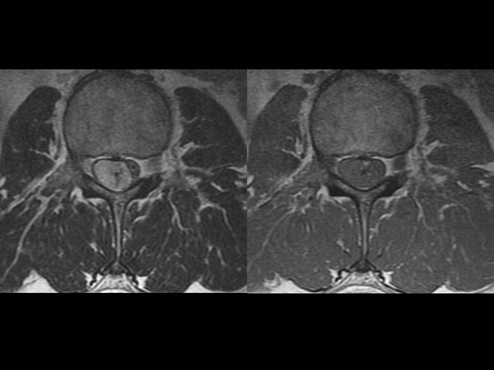
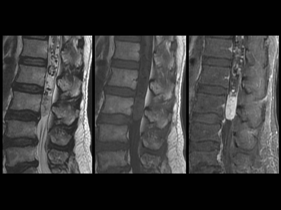
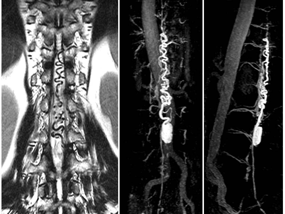
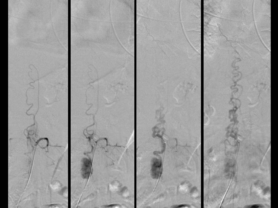
| Discussion | 정답: Spinal intradural-extramedullary hemangioblastoma (with AV shunt) 설명: L3 level의 thecal sac 내부에 약 2.6cm 크기의 T1-isointense, T2-hyperintense, homogenous strong enhancement를 보이는 mass가 있음. Mass의 내부에 flow void들이 있고 주변으로도 perimedullary vessel의 engorgement에 signal void가 있음. MRA에서 tumor와 연결되는 engorged vascular structure들이 보임. L2 segmental artery에서 origin하는 radioculopial artery가 tumor를 supply하고 있으며 tumor staining과 동시에 perimedullary vein들로 blood가 shunting되고 있어 arteriovenous shunting을 동반하고 있을 것으로 보임. 고찰: Most spinal hemangioblastomas present intramedullary growth, and are the third general cause of intramedullary tumors. Small and clinically nonsignificant extramedullary hemangioblastomas are well documented in the literature and are generally observed adherent to the cauda equina, filum terminale or anterior and posterior roots of the spinal nerves. Accoding to an extensive review of the literature covering 138 cases, found 38 cases of intradural extramedullary hemangioblastoma, of which 28 were located on nerve roots (including the cauda equina) and 10 cases were extramedullary tumors, as in the current report, but these occurred in patients that presented signs of VHL or were asymptomatic and showed incidental findings at autopsy. Hemangioblastomas usually show enlargement of the spinal cord associated with a cyst. Contrast-enhanced images can show one or more intensely enhancing nodules, which may be in the cyst wall or separate. Spinal cord hemangioblastomas larger than 2.4 cm are often accompanied by vascular flow voids, in contrast to homogeneous enhancement of smaller hemangioblastomas. The presence of an enlarged dorsal draining pial vein, seen as serpiginous flow voids on MR image, was helpful in the diagnosis. These findings strongly suggested hemangioblastoma. The most important differential diagnosis is a nerve root sheath tumor. But this can be ruled out with the findings of abnormal vascular structure. 참고문헌: 1. Barbosa-Silva E, Carvalho GT, Frota Mde O, Sousa AA, Souza CB. Intradural extramedullary hemangioblastoma. Arq Neuropsiquiatr. 2009 Jun;67(2B):530-3. 2. Chen CY, Chen PH, Yao MS, Chu JS, Chan WP. MRI of hemangioblastoma in the conus medullaris. Comput Med Imaging Graph. 2008 Jan;32(1):78-81. Epub 2007 Oct 10. |
| Correct answer |
Correct AnswerSemi-Correct Answer |