HOME > Weekly Case > Weekly Case
Weekly Case
종결
Case No. 230
2010-11-23~2010-12-05
출제자 : 관리자
Hit : 377
| Period | 2010-11-23~2010-12-05 |
|---|---|
| Diagnosis | |
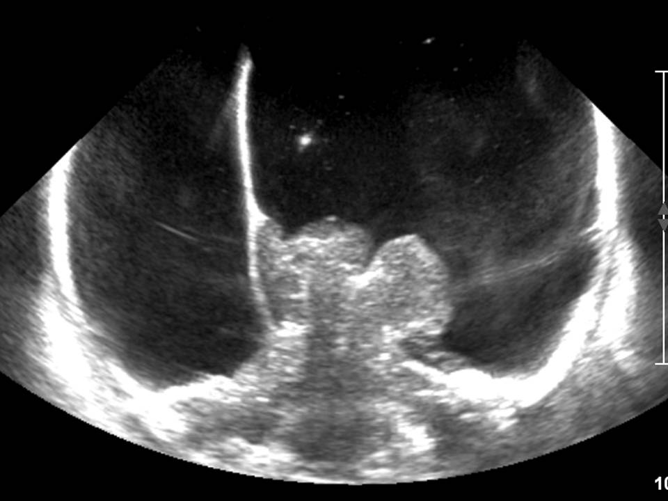
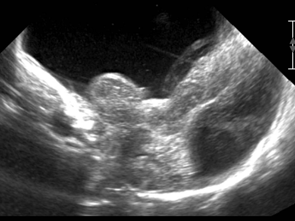
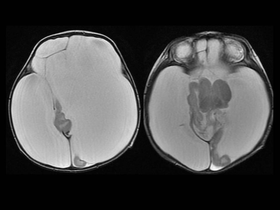
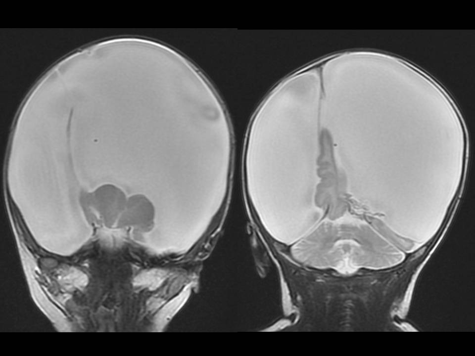
| Clinical information | F/0 C.C. : Anterior fontanelle bulging, seizure. |
| Discussion | 정답: Hydranencephaly 설명: Falx cerebri와 medial temporal lobe의 일부가 보임. Thalamus, brainstem, cerebellum은 비교적 정상적으로 보임. CT와 MRI에서는 thalamus, temporal lobe, occipital lobe의 일부, falx, brain stem, cerebellum이 보이나 나머지 cerebral parenchyma는 보이지 않고 CSF로 채워져 있음. 고찰: Hydranencephaly occurs in less than 1/10,000 births. Hydranencephaly has the appearance of near-total or total absence of that part of the brain supplied by anterior and middle cerebral arteries. The parts of the brain supplied by the posterior circulation are preserved, so the posterior temporal lobes, occipital lobes, thalami, and intratentorial structures are well formed. Hydranencephaly should not be confused with hydrocephalus, which is an accumulation of CSF in the ventricles. CT and MRI can demonstrate absence of most of supratentorial structures, with preservation of falx, thalami, and various amounts of the occipital lobes and basal ganglia. When fetal USG is performed in early gestation, hydrocephalus and alobar holoprosencephaly can be difficult to distinguish from hydranencephaly. Hydranencephaly has guite poor prognosis, and the patient die in 1st year of life. 참고문헌: David M. Yousem, Robert I. Grossman, The Requisites Neuroradiology 2010: 284 |
| Correct answer |
Correct AnswerSemi-Correct Answer |