HOME > Weekly Case > Weekly Case
Weekly Case
종결
Case No. 217
2010-07-06~2010-07-18
출제자 : 관리자
Hit : 387
| Period | 2010-07-06~2010-07-18 |
|---|---|
| Diagnosis | |
| Clinical information | 58/M C.C: Sudden onset of neck pain and tingling sensation of the upper extremities |
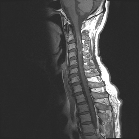
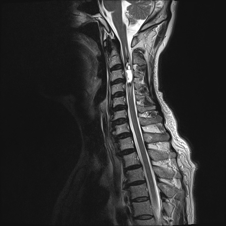
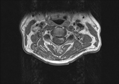
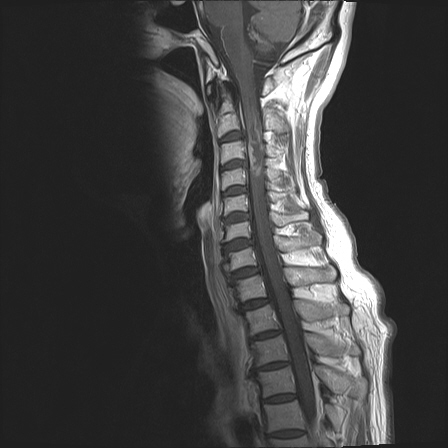
| Discussion | Diagnosis : spinal cord ependymoma Imaging finding A relatively well-demarcated intradural, intramedullary mass with cystic change is demonstrated at the C3 level of the spinal cord on the sagittal and axial cervical MR images. The mass is hyperintense on the sagittal T2WI with intratumoral cystic change. Irregular dark signal rim is seen at the upper and lower margin of the mass on T2WI, suggesting hemorrhage. Mild syrinx formation is associated at the cranial segment of the affected spinal cord. Peripheral nodular enhancement is also seen, after gadolinium enhancement. Review Spinal ependymomas arise from ependymal cells lining the central canal or its remnants and from the cells of the ventriculus terminalis in the film terminale. Ependymomas occur most often in the conus medullaris and film terminale. Myxopapillary ependymoma is most commonly found in this location. Cervical cord is the most common site for intramedullary ependymomas (usually cellular or mixed ependymomas). MR imaging shows a widened cord or filum terminale mass. Most ependymomas are isointense compared to cord on T1WI. Typically hyperintense on T2WI, and heterogeneous signal intensity when intratumoral cyst formation is present. Hypointensity at the tumor margin on T2WI is common in intramedullary ependymomas and is suggestive of ependymomas, but not pathognomic. Tumor-related syringes are more common in ependymomas, than astrocytomas. Ependymomas enhance strongly following contrast adminstration. The tumor margin is clearly delineated on contrast enhanced scans. Reference 1. Anne G. Osborn. Diagnostic neuroradiology. Mosby, St. Luis, 1994, pp. 906-909 2. Sub B, Wang CC, Wang J, Liu A. MRI features of intramedullary spinal cord ependymomas. Journal of neuroradiology 2006; 13:346-351 |
| Correct answer |
Correct AnswerSemi-Correct Answer |