HOME > Weekly Case > Weekly Case
Weekly Case
종결
Case No. 216
2010-06-28~2010-07-11
출제자 : 관리자
Hit : 384
| Period | 2010-06-28~2010-07-11 |
|---|---|
| Diagnosis | |
| Clinical information | 45/F C.C.: Right hearing disturbance and tinnitus |
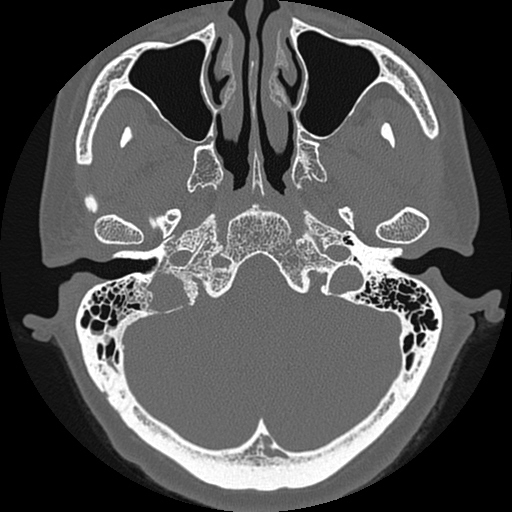
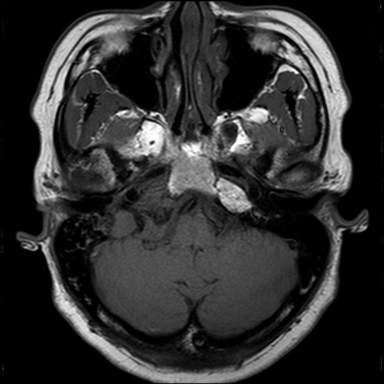
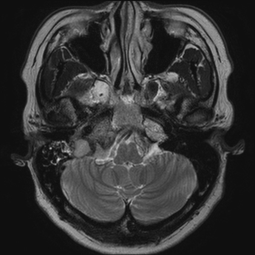
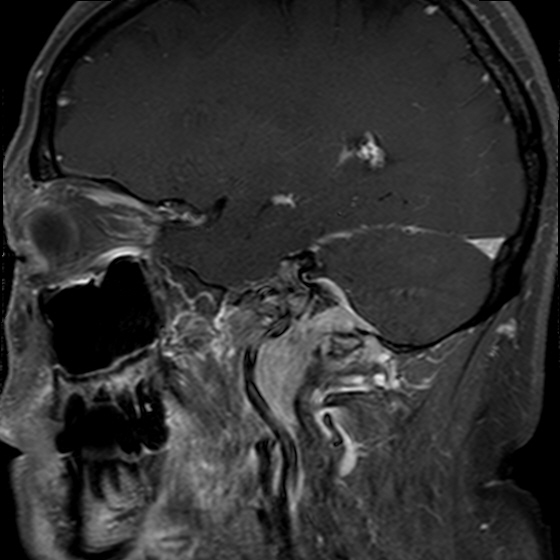
| Discussion | Diagnosis : Jugular foramen meningioma Imaging finding Axial temporal bone CT shows diffuse permeative bone destruction of the right jugular foramen and the right petrous bone, with relative preservation of bone density and architecture. T1- and T2WIs of the right temporal bone demonstrate an isointense solid mass infiltrating the right jugular foramen, the ipsilateral side of the clivus and the middle ear cavity. The mass is also encasing the right distal ICA. Note the thick enhancing dura on the surface of the homogeneously enhancing mass suggesting the dural tail sign on the gadolinium-enhanced sagittal T1WI. Review Primary jugular foramen meningioma is a common tumor in an unusual location. Meningiomas arising primarily in the jugular foramen are characterized by diffuse skull base infiltration. This locally invasive tendency differentiates primary jugular foramen meningioma from meningioma that involves the jugular foramen secondarily. On MRI, jugular foramen meningiomas appear iso- to hypointense in signal intensity on T1WIs and intermediate in signal intensity on T2WIs, without flow voids. Intense uniform enhancement is a typical finding after contrast administration. On CT, jugular foramen meningiomas usually have a permeative, irregular appearance to the bone margins of the jugular foramen and surrounding skull base, with relative preservation of bone density and architecture. The most common differential possibilities include paragangliomas and schwannomas. Paragangliomas are characterized by aggressive permeative-destructive bone change on CT. Other helpful differentiating features of paraganglioma include the presence of flow voids within the mass, best demonstrated on precontrast T1WIs, and, less commonly, hyperintense foci on the precontrast T1WIs representing foci of hemorrhage or slow flow within the mass (“salt and pepper appearance”). Angiography shows a highly vascular mass, typically demonstrating early arteriovenous shunting. Differential characteristics of schwannoma include a well-defined, smooth bony margin on CT as well as marked T2 hyperintensity on MRI, a well-circumscribed, elongated configuration following the course of the cranial nerves, and occasional cystic degeneration. Reference 1. Macdonald AJ, Salzman KL, Harnsberger HR, Gilbert E, Shelton C. Primary jugular foramen meningioma: Imaging appearance and differentiating features. AJR 2004; 182: 373-7. 2. Gilbert ME, Shelton C, Macdonald A, Salzman KL, Harnsberger HR, Sharma PK et al. Meningioma of the jugular foramen: Glomus jugulare mimic and surgical challenge. Laryngoscope 2004; 114: 25-32. |
| Correct answer |
Correct AnswerSemi-Correct Answer |