HOME > Weekly Case > Weekly Case
Weekly Case
종결
Case No. 215
2010-06-21~2010-07-04
출제자 : 관리자
Hit : 383
| Period | 2010-06-21~2010-07-04 |
|---|---|
| Diagnosis | |
| Clinical information | M/54 C.C.: Progressive quadriplegia, mental change |
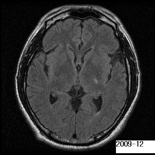
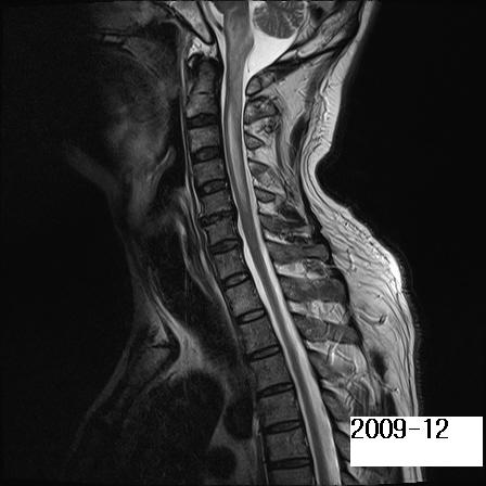
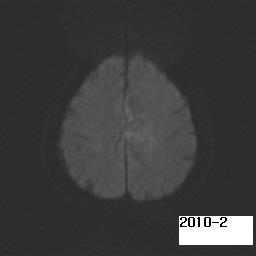
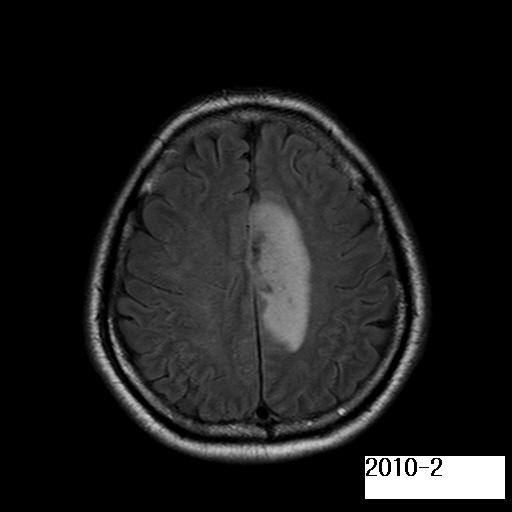
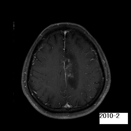
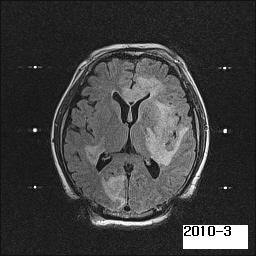
| Discussion | Diagnosis ; Intravascular large B-cell lymphoma. Imaging finding The initial sagittal FLAIR image shows diffuse ill-defined hyperintense lesion with swelling involving the cervical spinal cord and the medulla. There is also noted a tiny focal hyperintense lesion at the posterior limb of the internal capsule on the axial FLAIR image, which was considered non-specific. Despite of steroid therapy under the impression of acute transverse myelitis, quadriplegia was aggravated with mental deterioration. The F/U brain MRI after 2-month demonstrate a newly detected hyperintense lesion in the left cingulate gyrus and corona radiata on the axial FLAIR image with mild irregular enhancement, of which diffusion is mildly restricted. Axial FLAIR image performed after 1-month for stereotactic biopsy demonstrates marked progression of the extent of hyperintensities involving thecorpus callosum, the left basal ganglia and the right occipital lobe. Stereotatic biosy of brain lesion revealed intravascular large B-cell lymphoma. The patient died 3 month after the onset of symptom. Review Intravascular large B-cell lymphoma is a rare disease entity with a high incidence of CNS involvement at diagnosis. This is a rapidly progressive and disseminated, often fatal, aggressive lymphoma, characterized by selective growth of lymphoma cells only in the lumina of small vessels in various organs. Clinical features of spinal cord involvement are not uncommon. Because no specific clinical or laboratory findings are associated with intravascular lymphomatosis, the diagnosis is rarely established before histologic examination during autopsy. MRI findings in intravascular lymphomatosis include high-signal deep white matter lesions and infarct like, high-signal lesions in vascular territories on T2-weighted images. After the administration of contrast material, enhancement can be masslike. Various patterns of parenchymal and meningeal enhancement may also be seen. Differential diagnoses is vasculitis, demyelinating disease, classical primary cerebral lymphoma and gliomatosis cerebri. Reference 1. Shimada K, Murase T, Matsue K, Okamoto M, Ichikawa N et al. Central nervous system involvement in intravascular large B-cell lymphoma: A retrospective analysis of 109 patients. Cancer Sci. 2010 Mar 6. 2. Martin-Duverneuil N, Mokhtari K, Behin A, Lafitte F, Hoang-Xuan K, Chiras J. Intravascular malignant lymphomatosis Neuroradiology. 2002;44(9):749-54. 3. H. Wayne Slone, Joseph J. Blake, Rajul Shah, Sangeeta Guttikonda, and Eric C. Bourekas CT and MRI Findings of Intracranial Lymphoma AJR 2005; 184: 1679 - 1685. |
| Correct answer |
Correct AnswerSemi-Correct Answer |