HOME > Weekly Case > Weekly Case
Weekly Case
종결
Case No. 214
2010-06-14~2010-06-27
출제자 : 관리자
Hit : 356
| Period | 2010-06-14~2010-06-27 |
|---|---|
| Diagnosis | |
| Clinical information | 10/M C.C.: exotropia of the left eye |
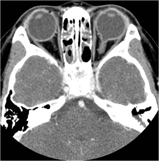
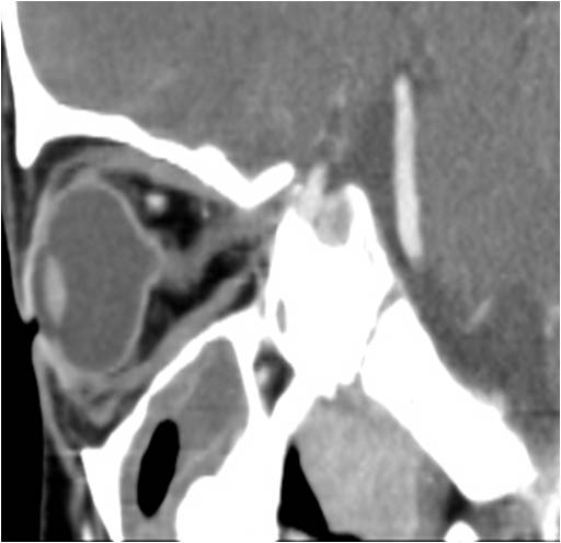
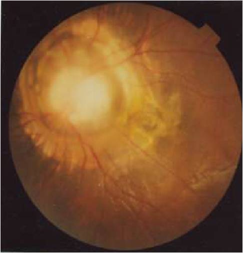
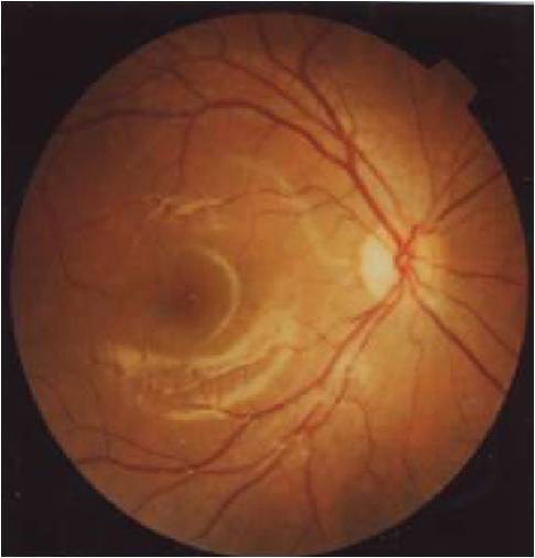
| Discussion | Diagnosis : Coloboma Imaging finding Enhanced axial CT scan shows a small focal defect with outpouching of vitreous in the left posterior globe. No other retrobulbar colobomatous cyst or other abnormal enhancement within the defect was noted. Enhanced sagittal CT scan reveals a dehiscence of posterior globe through a defect centered at the location of the optic nerve head. Contrary to the normal fundoscopic finding on the right side, there is an enlarged excavated optic disc which resembles morning glory flower. Also noted is loss of optic disc neuroretinal rim with chorioretinal atrophy involving the macula. Review A coloboma is a notch, gap, hole or fissure that is congenital or acquired and in which a tissue or portion of a tissue is lacking. Ocular coloboma may involve the iris, lens, ciliary body, retina, choroid, optic nerve, or sclera. Colobomas are usually transmitted as an autosomal dominant trait and are bilateral in 60 % of patients. Ophthalmoscopically, optic nerve coloboma characteristically shows enlargement of the papillary areas, with partial or total excavation of the disc. The most common symptoms are decreased visual acuity. The visual acuity in eyes with optic nerve colobomas is variable and may range from normal to abnormal light perception. The only predictive factor of visual acuity in a patient with optic disc coloboma is the degree of foveal involvement by the coloboma. Strabismus and nystagmus secondary to poor visual acuity can also develop. A coloboma can form in normal-sized eyes as well as microphthalmic eyes. In a small percentage of microphthalmic eyes with coloboma, a defect in the sclera allows an extraocular herniation of the intraocular neural ectoderm to form an orbital cyst with a tunnel-like connection to the globe, which is called a colobomatous cyst. Morning glory anomaly is a form of dysplastic colobma. It is an anomaly of optic nerve head which is similar to morning glory flower in appearance. The CT and MR appearance of optic disc coloboma includes a posterior global defect with optic disc excavation. The imaging appearance of morning glory anomaly is a large funnel-shaped disc excavation. The typical colobomatous cyst associated with coloboma of the optic nerve head is identified as a cyst extending posterior to orbit with microphtalmic eye on cross sectional imaging. |
| Correct answer |
Correct AnswerSemi-Correct Answer |