HOME > Weekly Case > Weekly Case
Weekly Case
종결
Case No. 211
2010-05-10~2010-05-23
출제자 : 관리자
Hit : 461
| Period | 2010-05-10~2010-05-23 |
|---|---|
| Diagnosis | |
| Clinical information | 50세 남자, seizure |
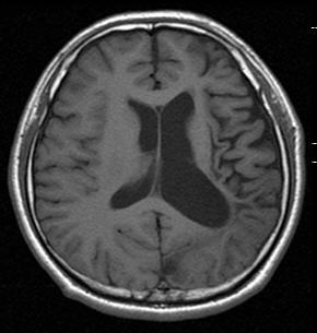
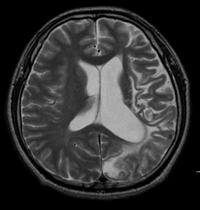
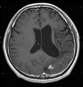
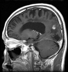
| Discussion | Dx. Cerebral sparganosis Cerebral sparganosis, caused by a migrating tape worm larva (spanganum) of the genus Spirometra, is a rare parasitic disease producing longstanding inflammation of brain tissue. Sparganosis has been reported to occur worldwide, but it is most common in East Asia. The major presenting symptoms of cerebral spanganosis are seizure, hemipanesis, and headache, with a chronic course. The primary characteristic of cerebral sparganosis is that the live worm migrates with undulating motion. The migrating worm makes a tunnel along the track of motion. A tunnel sign was conspicuously seen on postcontrast sagittal and coronal MR images as fusiform shaped, appearing hypointense on T1-weighted images, slightly hyperintense or isointense on T2-weighted images, and column or fusiform shaped on postcontrast MR images. The column-shaped or fusiform shaped tunnel showed a solid or hollow appearance with a smooth, well-defined margin. From a pathologic standpoint, the enhancing tunnels were the reactive inflammatory tissue or granuloma enwrapping the worm. The second characteristic of cerebral sparganosis is the conglomerated ring or bead-shaped enhancement, which represents an inflammatory granuloma. The wall of the ring-shaped granulomas is hypointense on T1-weighted images and isointense or slightly hypointense on T2-weighted images. The cause of the hypointensity or isointensity of the wall of the ring on T2-weighted images may be the same as those of a pyogenic abscess that presumably resulted from the free radicals produced by macrophages. The third characteristic of cerebral sparganosis is the alternate change of varying stages in the same image because of the long course of the disease, from the initial attack to admission to the hospital. Small, punctuate calcifications were around or in the degenerated or dead worm produced by deposition of calcareous corpuscles (particles containing calcifications). The calcifications could be clearly revealed on CT images but were obscure on MR images because of the insensitivity to calcifications. Meanwhile, unilateral ventricular dilation and focal cerebral cortical atrophy hinting the chronic stage may be caused by long-standing injuries by the worm. Cortical atrophy and bead-shaped enhancement denoting the chronic and acute phases, respectively, usually coexisted in the same lesion. References 1. Moon WK, Chang KH, Cho SY, et al. Cerebral sparganosis: MR imaging versus CT features. Radiology 1993;188:751-757. 2. Song T, Wang WS, Zhou BR, et al. CT and MR characteristics of cerebral sparganosis. Am J Neuroradiol 2007;28:1700-1705. |
| Correct answer |
Correct AnswerSemi-Correct Answer |