HOME > Weekly Case > Weekly Case
Weekly Case
종결
Case No. 210
2010-05-04~2010-05-16
출제자 : 관리자
Hit : 419
| Period | 2010-05-04~2010-05-16 |
|---|---|
| Diagnosis | |
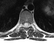
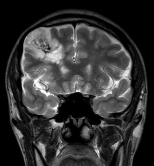
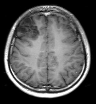
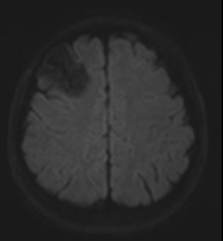
| Clinical information | 27세 남자, seizure for 20 years |
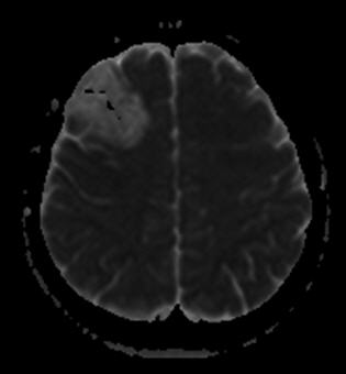
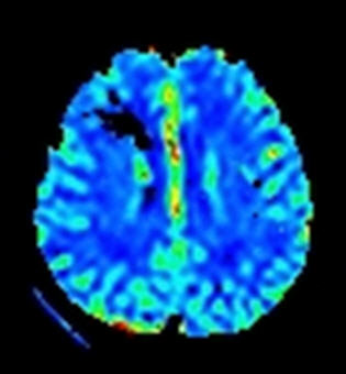
| Discussion | 진단명: Dysembryoplastic neuroepithelial tumor (DNET) Dysembryoplastic neuroepithelial tumor (DNET) is a rare benign, usually supratentorial, mixed neuronal-glial neoplasm characterized by multinodular architecture and a predominantly intracortical location. A background of cortical dysplasia adjacent to a DNET is often noted. A large majority of DNETs are located in the cortical region of the temporal lobe, with common extension into adjacent subcortical white matter and preferential involvement of the mesial structures. Patients are usually presented with intractable seizures. At CT, it appears as a moderately or markedly hypoattenuating cystlike lesion. Calcification is reported in less than 20% of cases. On MR images, it has a well-demarcated, multilobulated or gyriform appearance and is hypointense on T1-weighted images and markedly hyperintense on T2-weighted images. Yamasaki et al. reported that the ADC of DNET was higher than that of other WHO grade 1/2 glioma and other glioneuronal tumors due to the presence of large extracellular spaces and their low cellularity. There is a high frequency of bone remodeling of the adjacent calvaria at CT or MR imaging. Typically, no surrounding vasogenic edema is present, and enhancement at CT or MR imaging occurs in about one-third of cases but is not a prominent feature. Differential diagnoses include low-grade astrocytoma, ganglioglioma and oligodendroglioma. References 1. Shin JH, Lee HK, Khang SK, et al. Neuronal tumors of the central nervous system: radiologic findings and pathologic correlation. Radiographics 2002;22:1177-1189. 2. Kuroiwa T, Bergey GK, Rothman MI, et al. Radiologic appearance of the dysembryoplastic neuroepithelial tumor. Radiology 1995;197:233-238. 3. Yamasaki F, Kurisu K, Satoh K, et al. Apparent diffusion coefficient of human brain tumors at MR imaging. Radiology 2005; 235: 985-991. |
| Correct answer |
Correct AnswerSemi-Correct Answer |