HOME > Weekly Case > Weekly Case
Weekly Case
종결
Case No. 209
2010-04-26~2010-05-10
출제자 : 관리자
Hit : 380
| Period | 2010-04-26~2010-05-10 |
|---|---|
| Diagnosis | |
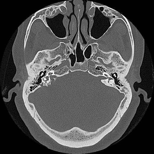
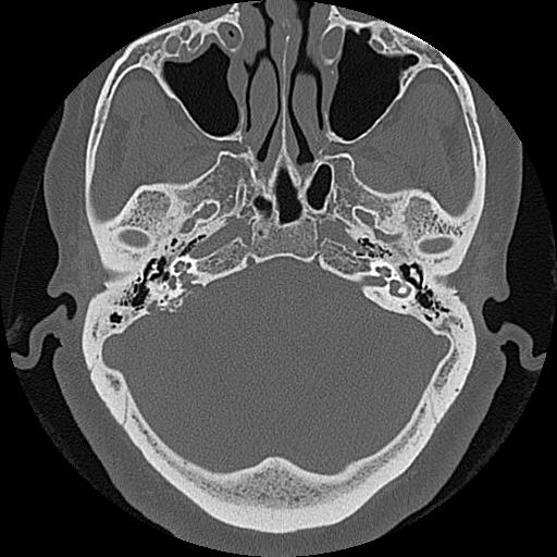
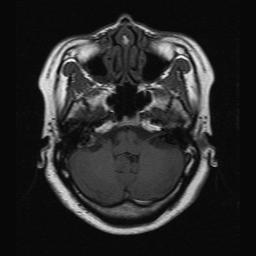
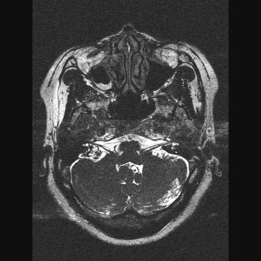
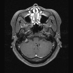
| Clinical information | 52/F Spinning sensation for 10 days, several attack |
| Discussion | 진단 : Endolymphatic sac tumor Imaging findings: Distinctive retrolabyrithine location; posteromedial surface of the petrous bone at the level of the vestibular aqueduct and endolymphatic sac, anteromedial to the sigmoid sinus CT; aggressive soft tissue mass with bony destruction, central speculated calcification with tumor matrix, MRI; hyperintense foci within tumor on T1WI, heterogenous enhancement on the T1-Gd images 고찰: Endolymphatic sac tumor Papillary adenocarcinoma of the temporal bone originate in the endolymphatic sac CT ; early lesion - a soft tissue mass with bone destruction is typically centered on the retrolabyrithine region, between the sigmoid sinus and internal auditory meatus at the site of the endolymphatic sac advanced lesion – spread to the adjacent structures, involve the middle ear, CPA cistern, jugular foramen, intratumoral bony fragments are frequently present in the endolymphatic sac tumors and a thin rim of bone may be seen MR; High SI is seen on both T1 and T2-weighted images. In small endolymphatic sac tumors increased signal intensity is usually present as a rim while in more advanced lesions multiple foci are distributed throughout the tumor. This high SI correlates with regions of subacute hemorrhage, proteinaceous cystic areas and cholesterol clefts found on histological examination. Intense enhancement in tumor on both CT and MR and angiographically these lesions may be highly vascular. Feeding vessels initially arise from branches of the ECA, most commonly the ascending pharyngeal artery. Preoperative embolization of tumor maybe of benefit by reducing operative blood loss. The development of bilateral tumors associated with von Hippel-Lindau disease, the incidence of endolymphatic sac tumors in this disease estimated 11-15%. |
| Correct answer |
Correct AnswerSemi-Correct Answer |