HOME > Weekly Case > Weekly Case
Weekly Case
종결
Case No. 207
2010-04-12~2010-04-25
출제자 : 관리자
Hit : 445
| Period | 2010-04-12~2010-04-25 |
|---|---|
| Diagnosis | |
| Clinical information | 35/m, neck pain, right hand numbness |
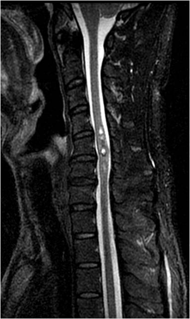
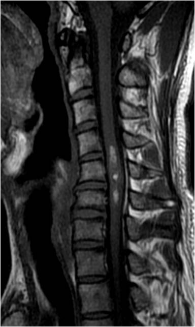
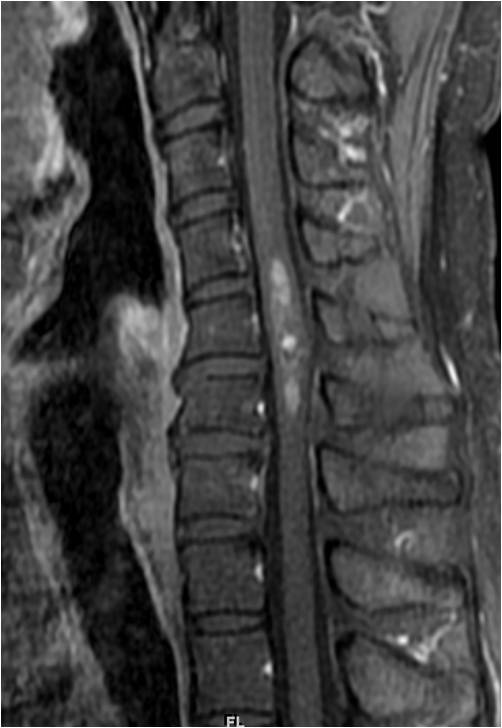
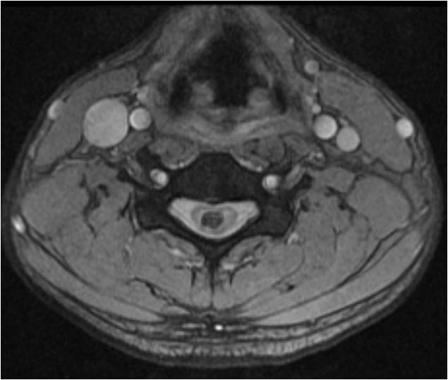
| Discussion | Cavernous malformation, spinal cord Imaging findings C4-6 level의 spinal cord 에 hemorrage-fluid level과 hemosiderin rim을 동반한 T2 강조 영상에서 고신호 강도의 cord lesion이 보이는 데, 조영 증강은 거의 보이지 않는다. 1. Definition: vascular lesion with lobulated, thin sinusoidal vascular channels, no interspersed neural tissue 2. Locules of blood with fluid-fluid lebels srrounded by very T2 hypointense rim 3. MR -T1: heterogeneous -T2: heterogeneous, hypointense rim(hemosiderin), no edema unless recent hemorrhage -T2*GRE: prominent blooming due to susceptibility effects -T1C+: enhancement absent/minimal 4. Clinical ourse varies from slow progression to acute quadriplegia -bleeding rate: 1-5% per lesion per year 5. DDx: intramedullary tumor, especially ependymoma |
| Correct answer |
Correct AnswerSemi-Correct Answer |