HOME > Weekly Case > Weekly Case
Weekly Case
종결
Case No. 206
2010-04-05~2010-04-18
출제자 : 관리자
Hit : 448

| Period | 2010-04-05~2010-04-18 |
|---|---|
| Diagnosis | |
| Clinical information | 48 year-old women with DM and CRF was performed hemodialysis. She was complained insonmia, acalcuria, and showed confusional mental status. |
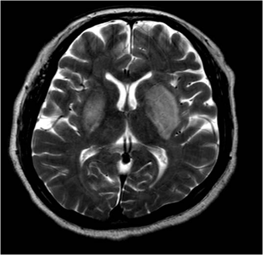
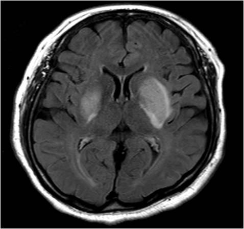
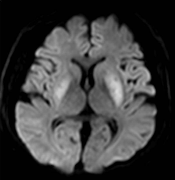
| Discussion | Extrapontine myelinolysis or osmotic demyelination syndrome Imaging findings 양측 basal ganglia는 edematous swelling을 보이고, 이 병변은 확산 강조 영상에서 고신호 강도를 보이며 ADC map에서 다양한 정도의 diffusion restriction을 보인다. Osmotic demyelniation syndrome: central pontine myelinolysis and/or extrapontine myelinolysis 1.Definition: aute demyelination caused by rapid shifts in serum osmolality -Classic setting: rapid correction of hyponatremia -But can occur in normonatremic patients -EPM can occur without CPM. 2. Location -CPM: 50% - EPM: 50% -Basal ganglia, cerebral white matter 3. MR -T2W1: confluent hyperintensity in central ponswith sparing of periphery and corticospinal tracts Symmetric hyperintensity in BG, WM (EPM) -DWI: restricted (mildly hyperintense) -ADC: variable, normal to mildly hyperintense |
| Correct answer |
Correct AnswerSemi-Correct Answer |