HOME > Weekly Case > Weekly Case
Weekly Case
종결
Case No. 204
2010-03-22~2010-04-04
출제자 : 관리자
Hit : 507
| Period | 2010-03-22~2010-04-04 |
|---|---|
| Diagnosis | |
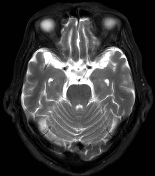
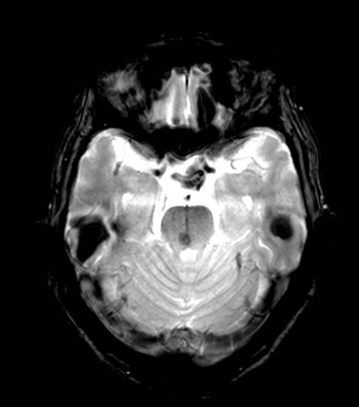
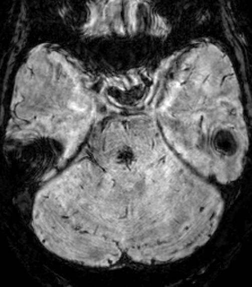
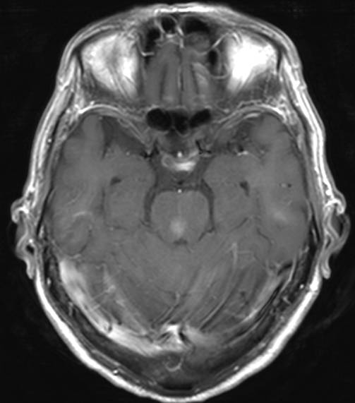
| Clinical information | To work up for brain metastasis in a 75 year-old man with lung cancer |
| Discussion | 정답: Capillary telangiectasia Imaging findings: Pons의 뒤쪽으로 조영 증강영상에서는 faint brush-like enhancement가 보이지만, T2 강조 영상에서 병변은 보이지 않는다. 이 병변은 T2*GRE 영상에서 약간 낮은 신호 강도로 보이는데, SWI (Susceptibility-weighted image) 에서 매우 dark signal intensity로 보인다. 또한 앞쪽으로 아마도 collecting vein에 의한 linear dark signal intensity가 보인다. Definition: clusters of capillaries interspersed with normal brain parenchyma T2WI: 50% normal; 50% stippled foci of hyperintensity T2*GRE: lesion moderately but not profoundly hypointense due to slow blood flow with oxy to deoxyhemoglobin. T1C+: faint stippled or speckled brush-like enhacement, may show punctuate, linear/branching vessles with/without collecting vein DDx 1. Metastasis: very strong enhancement > faint enhancement 2. cavernous malformation: usually not enhancement |
| Correct answer |
Correct AnswerSemi-Correct Answer |