HOME > Weekly Case > Weekly Case
Weekly Case
종결
Case No. 203
2010-03-11~2010-03-21
출제자 : 관리자
Hit : 438
| Period | 2010-03-11~2010-03-21 |
|---|---|
| Diagnosis | |
| Clinical information | CC: Rt. Leg pain, 2 yrs, 78/female |
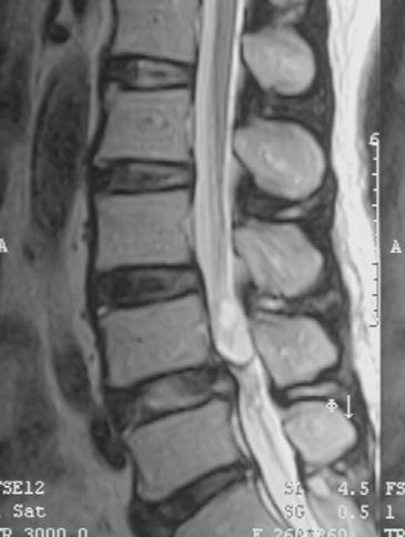
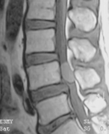
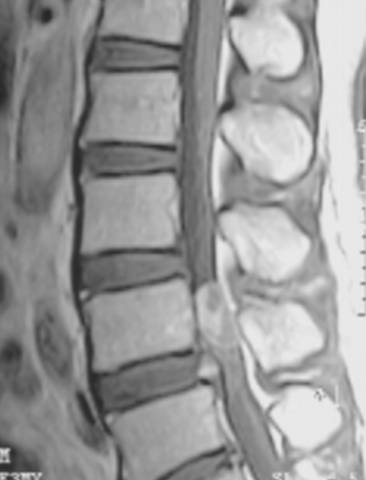
| Discussion | Diagnosis: paraganglioma Image finding: There is ovoid lesion with T2 high and T1 iso SIs in the intradural extramedullary lower L spine. The lesion shows heterogeneous enhancement. On T2WI, there is some dark SI on the periphery of the mass. Review) Although being nonspecific, paraganglioma has been reported to have the tendency to show dark peripheral T2 SI d/t hemosiderin or vascular voids. For differentiation, neurogenic tumor and ependymoma (myxopapillary type) should be considered. The meningioma is a less possible diagnosis because it shows relatively hyperintense T2 SI and rarely involves the lumbar spine. For rare condition as a well enhancing tumor in this region, capillary hemangioma and paraganglioma should be considered. Ref). Yang SY, Jin YJ, Park SH, Jahng TA, Kim HJ, Chung CK. Paragangliomas in the cauda equina region: clinicopathoradiologic findings in four cases. J Neurooncol. 2005 Mar;72(1):49-55. |
| Correct answer |
Correct AnswerSemi-Correct Answer |