HOME > Weekly Case > Weekly Case
Weekly Case
종결
Case No. 200
2010-01-11~2010-01-24
출제자 : 관리자
Hit : 414
| Period | 2010-01-11~2010-01-24 |
|---|---|
| Diagnosis | |
| Clinical information | Chief complaint: lt. ocular pain, 3 months, F/67 |
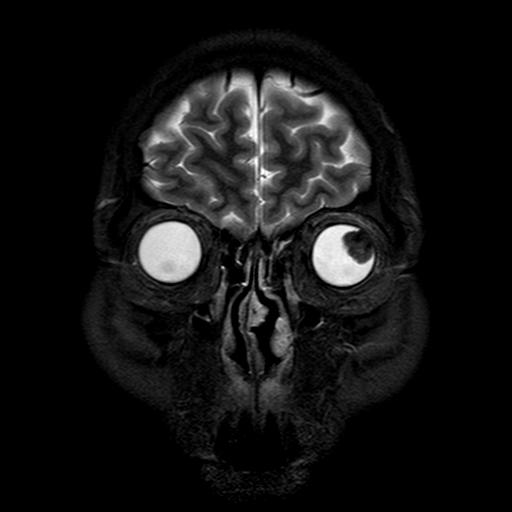
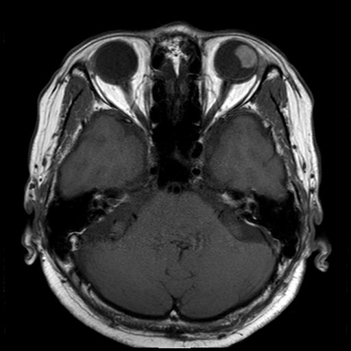
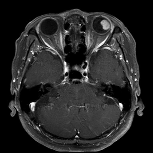
| Discussion | 진단명: ocular melanoma Imaging finding: T2WI reveals a lesion with dark SI in the lt. upper lateral eyeball. The lesion shows high SI on T1WI and homogeneous enhancement on enhanced T1WI. Review: CT findings - NECT: solid soft tissue density mass Calcification rare; may occur after therapy - DECT: diffuse moderate enhancement MR findings T1WI: mildly to strongly hyperintense T2WI: strongly hypointense T1 contrast enhacement: moderate, diffuse enhancement DDx chorioretinal or scleral inflammatory disease, benign chorioretinal masses (osteoma, hemangioma, hamartoma, angioma, telangiectasia), metastasis, retinoblastoma, chorioretinal detachment |
| Correct answer |
Correct AnswerSemi-Correct Answer |