HOME > Weekly Case > Weekly Case
Weekly Case
종결
Case No. 198
2009-11-27~2009-12-13
출제자 : 관리자
Hit : 470
| Period | 2009-11-27~2009-12-13 |
|---|---|
| Diagnosis | |
| Clinical information | 9/M, CC: Headache, ocular pain. |
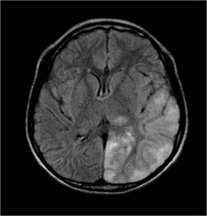
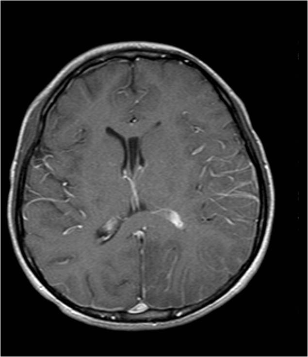
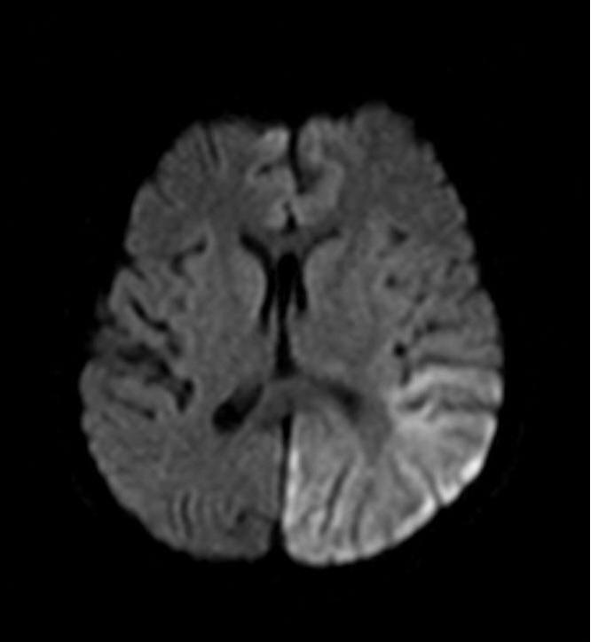
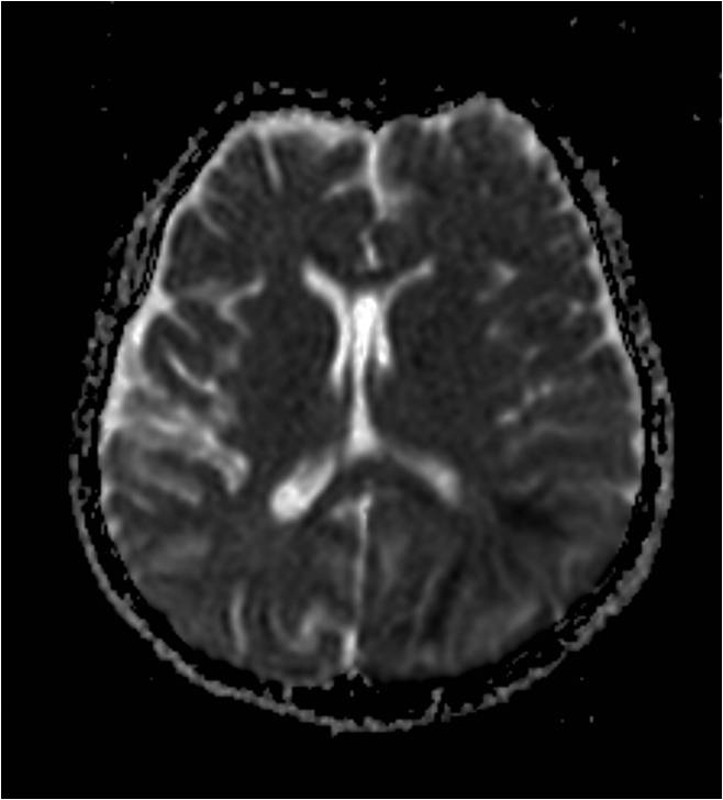
| Discussion | 최종진단: MELAS (mitochondrial myopathy, encephalopathy, lactic acidosis, and stroke-like episodes) syndrome Imaging findings: FLAIR영상에서 left thalamus와 temporooccipital lobe에 multiple hyperintense lesion들이 있음. DWI에서도 병변들은 고신호 강도를 보이고 ADC map image에서는 heterogeneous signal intensity를 보임. 조영증강 T1강조영상에서 병변의 뚜렷한 조영증강은 보이지 않음 고찰: MELAS syndrome •Inherited disorder of intracellular energy production caused by point mutation in mtDNA. •Acute: stroke-like cortical lesions -“Shifting spread” (appearance, disappearance, reappearance elsewhere) is classic. -Lesions cross typical vascular territories. •Most common signs/symptoms -Acute onset: headache, followed by hemianopsia, psychosis, aphasia -Other signs/symptoms: seizures, muscle weakness(myopathy), sensorioneural hearing loss •Age: onset of stroke-like episodes usually occurs in childhood, early adulthood (mean age onset = 15y) •MR Findings -Multiple strokelike lesions primarily in the parietal and occipital cortex -Subcortical white matter, BG, brain sten, and cerebellum may also be involved, not corresponding to major vascular territories -T2WI: high signal in subcortical WM, basal ganglia, cerebellum infarcts -T2GRE: no hemorrhage -DWI Acute: DWI positive ADC: variable but often normal or only slightly decreased. |
| Correct answer |
Correct AnswerSemi-Correct Answer |