HOME > Weekly Case > Weekly Case
Weekly Case
종결
Case No. 197
2009-11-09~2009-11-22
출제자 : 관리자
Hit : 487
| Period | 2009-11-09~2009-11-22 |
|---|---|
| Diagnosis | |
| Clinical information | 61/F, CC: Headache and left side weakness (1DA). |
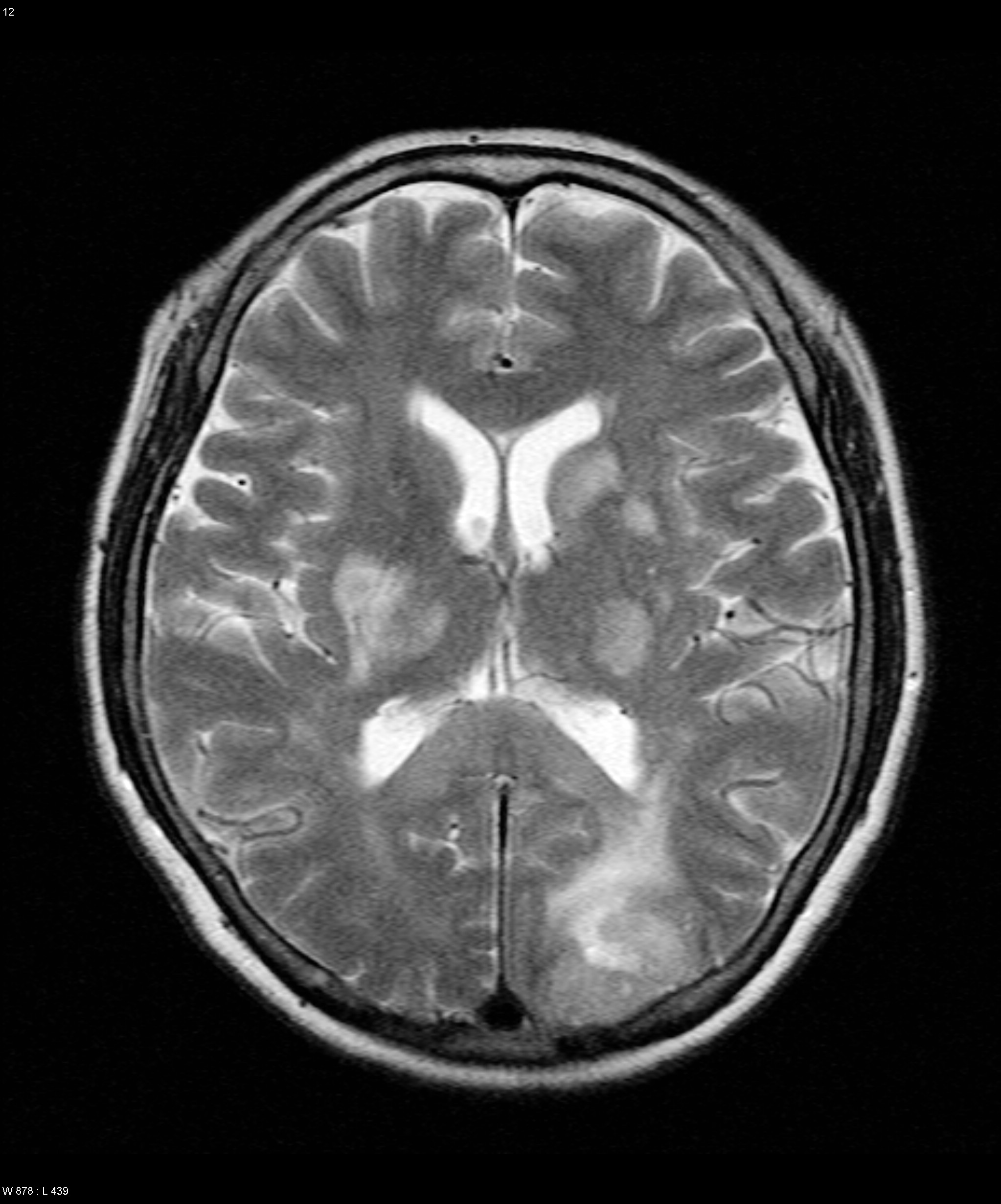
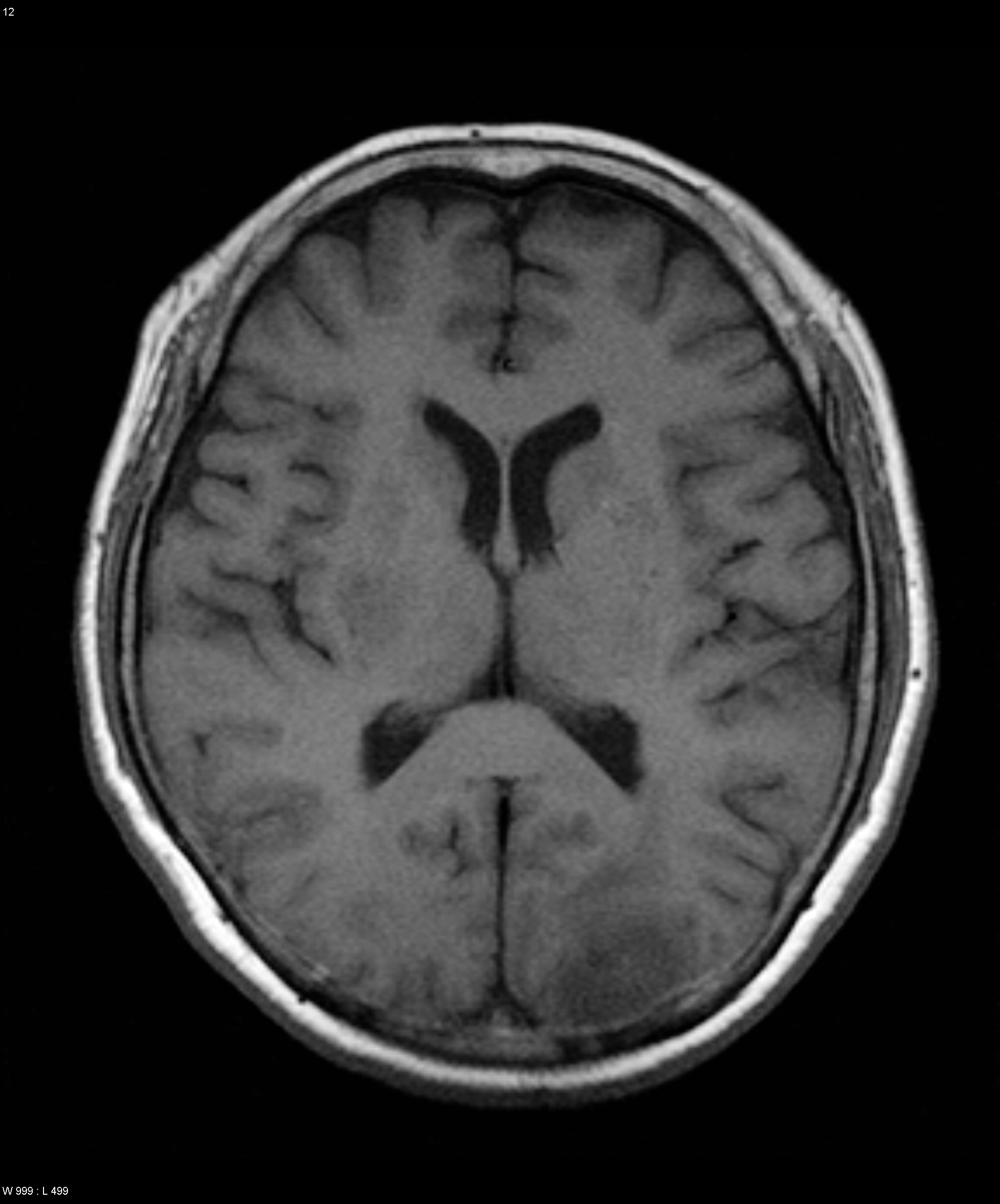
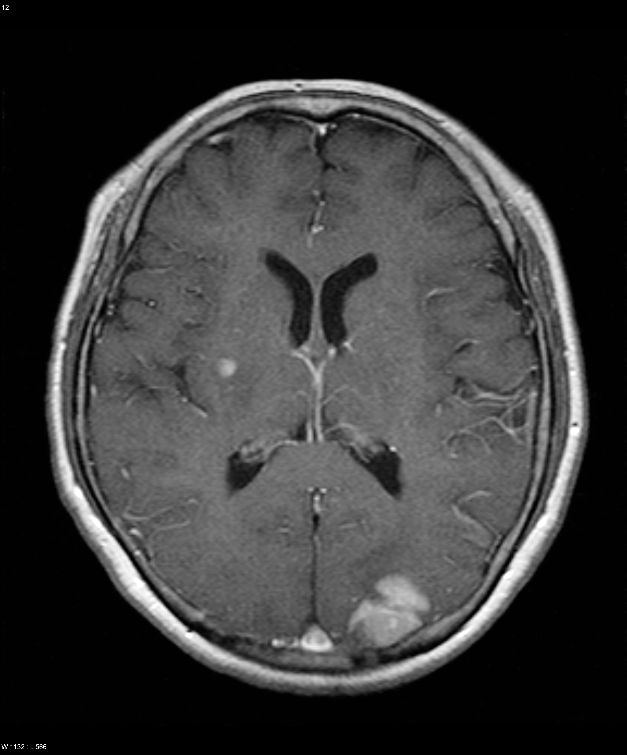
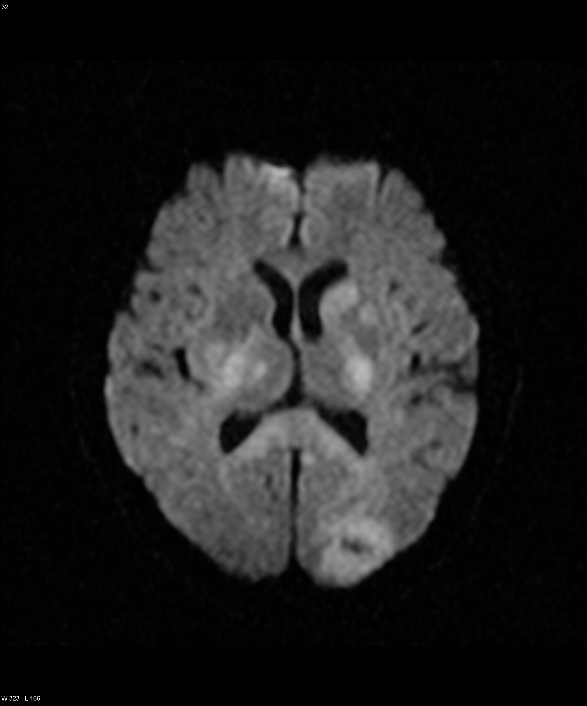
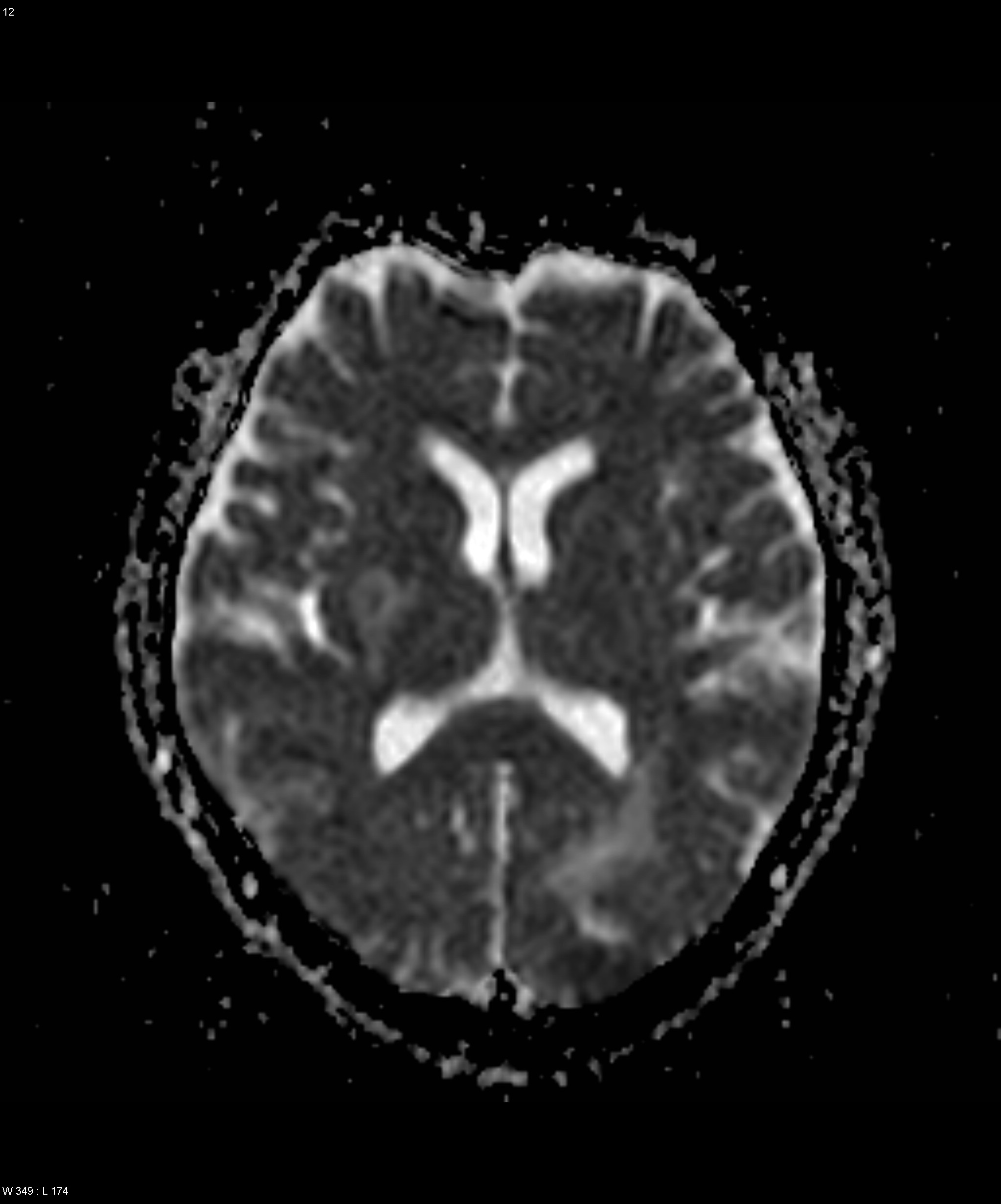
| Discussion | 최종진단: Lymphoma Imaging findings: T2강조영상에서 양측 basal ganglia, thalamus, corpus callosum의 splenial portion 및 left occipital lobe에 multiple hyperintense lesion들이 있음. T1강조영상에서 저신호 또는 동등 신호강도를 보이고, DWI에서는 고신호 강도를 보임. ADC map image에서는 heterogeneous signal intensity를 보임. 조영증강 T1강조영상에서 right basal ganglia와 left occipital lobe lesion들은 강한 조영증강을 보이나 그 외의 병변들은 조영증강되지 않음. 고찰: Primary CNS lymphoma Best diagnostic clue: Enhancing lesion(s) within basal ganglia, periventricular WM Location: 90% supratentorial - Deep gray nuclei commonly affected - Lesions cluster around ventricles, GM-WM junction - Often involve, cross corpus callosum - Frequently abut, extend along ependymal surfaces Morphology - Solitary mass or multiple lesions - May be circumscribed or infiltrative MR Findings - T1WI: isointense/ hypointense to cortex - T2WI: isointense/ hypointense to cortex - DWI: hyperintense with restricted diffusion - Enhanced T1WI: Immunocompetent: strong homogeneous Immunocompromised: peripheral enhancement with central necrosis or homogeneous |
| Correct answer |
Correct AnswerSemi-Correct Answer |