HOME > Weekly Case > Weekly Case
Weekly Case
종결
Case No. 194
2009-10-12~2009-10-26
출제자 : 관리자
Hit : 462
| Period | 2009-10-12~2009-10-26 |
|---|---|
| Diagnosis | |
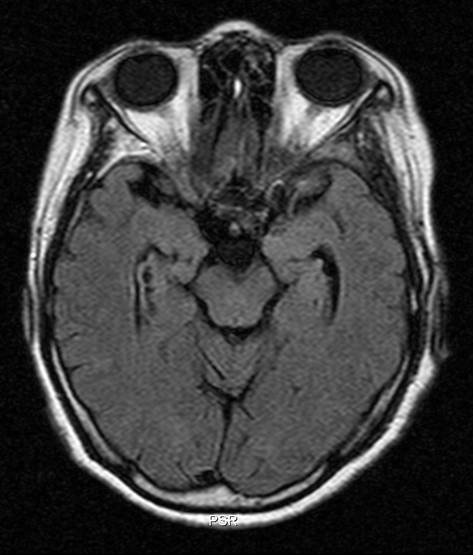
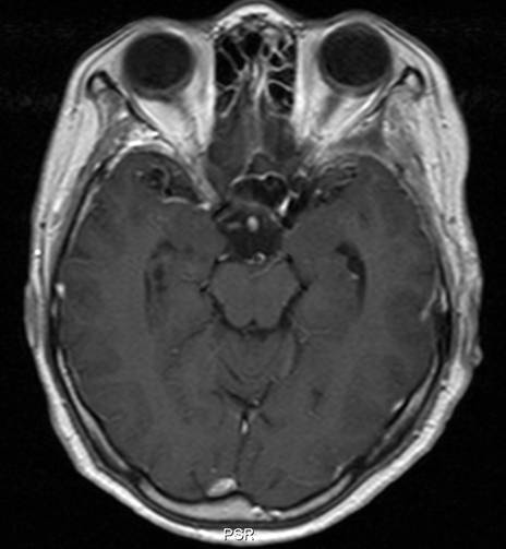
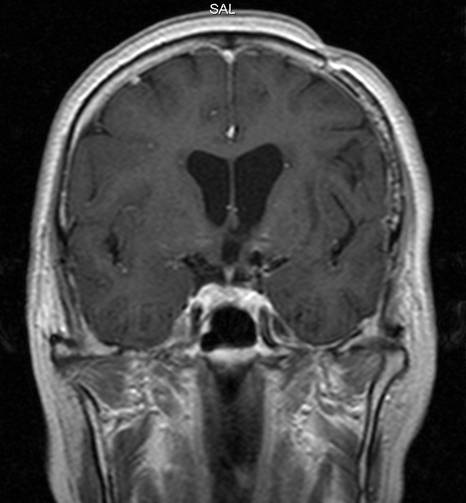
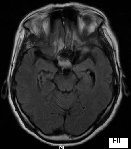
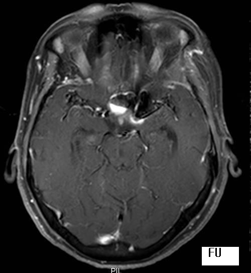
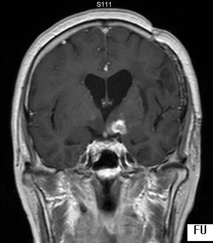
| Clinical information | 62 year old female with decreased visual acuity in the both eyes developed during several months and right side weakness of sudden onset. He underwent surgical clipping with wrapping of the aneurysms at Acom and left ICA bifurcation one year ago. |
| Discussion | Diagnosis: muslin-induced granuloma (Muslinoma) Findings In comparison with initial MRI obtained 3 months after clipping, 1-year follow-up MRI shows enhancing mass with thick enhancing rim surrounding the previously clipped aneurysm. Axial FLAIR image shows high signal intensity consistent with edema at subfrontal region and optic chiasm. Review Wrapping: reinforcement/wrapping of certain (anatomic configuration, location etc) IC aneurysm with muslin, cotton gauze in addition to clipping Muslinoma: FB inflammatory reaction, arachnoiditis/ inflammatory mass Sx: Visual loss (A-com); 1-24mo after surgery, Headache, Seizure, malaise, ischemic (vasculitis) MRI: rim enhancing collection/ relatively low SI on T2 Treatment: corticosteroid, immunosuppressant, surgical lysis of adhesions or observation Prognosis: unpredictable Reference Brochert, A. et al. MRI in a case of muslin-induced granuloma Neuroradiology 2003; 45: 82-84 |
| Correct answer |
Correct AnswerSemi-Correct Answer |