HOME > Weekly Case > Weekly Case
Weekly Case
종결
Case No. 193
2009-09-14~2009-09-27
출제자 : 관리자
Hit : 496
| Period | 2009-09-14~2009-09-27 |
|---|---|
| Diagnosis | |
| Clinical information | 42 year old male with AIDS with left side weakness. Known history of zoster infection, pulmonary tbc, oral candidiasis during recent 2 years. |
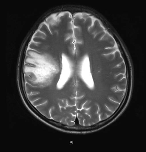
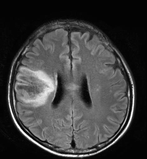
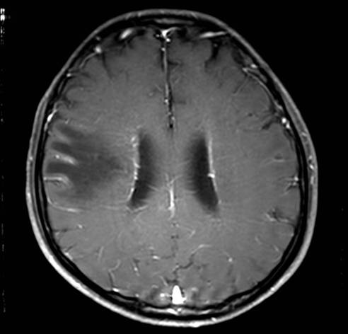
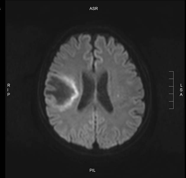
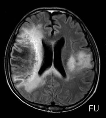
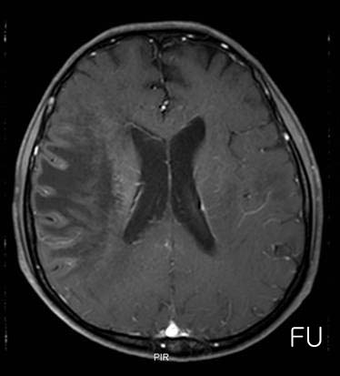
| Discussion | Diagnosis: Progressive Multifocal Leukoencephalopathy (PML) in HIV(+) patient Image Finding: Initial MRI shows coalescent abnormal FLAIR and T2 signal in the white matter of right periventricular and centrum semiovale areas with mild mass effect. Diffusion restriction along peripheral portion of the lesion. Lesions are low signal on T1. There is no abnormal contrast-enhancement after gadolinium injection. There is no significant mass-effect. F/U MRI after 3 months shows midline crossing extension of white matter lesion to left side and minimal contrast enhancement. Review: * Severe demyelinating disease of the CNS d/t JC polyoma virus infection of oligodendrocytes. * Develops in patients with impaired T-cell function as occurs in HIV, malignancy or immunosuppressive drugs users. * Symptoms include progressive focal and diffuse deficits (visual, motor,cognitive deficits and memory loss, personality change) * Leading to death a few months after diagnosis MRI findings 1. patchy, (usually) nonenhancing WM lesions, may be unilateral but ore often bilateral and asymmeteric 2. subcortical T2 hyper-/T1 iso- and later hypointensities, always affecting the U-fibers without mass effect. 3. frontal and parieto-occipital locations (m/c) 4. restricted diffusion of protons. References Bergui, M. et al. Progressive multifocal leukoencephalopathy: diffusion-weighted imaging and pathological correlations Neuroradiology (2004) 46: 22?5 Cosottini, M. et al. Diffusion-weighted imaging in patients with progressive multifocal leukoencephalopathy Eur Radiol (2008) 18: 1024?030 |
| Correct answer |
Correct AnswerSemi-Correct Answer |