HOME > Weekly Case > Weekly Case
Weekly Case
종결
Case No. 192
2009-08-31~2009-09-13
출제자 : 관리자
Hit : 536
| Period | 2009-08-31~2009-09-13 |
|---|---|
| Diagnosis | |
| Clinical information | 46세 여자, headache and visual disturbance |
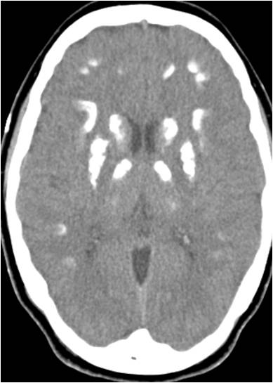
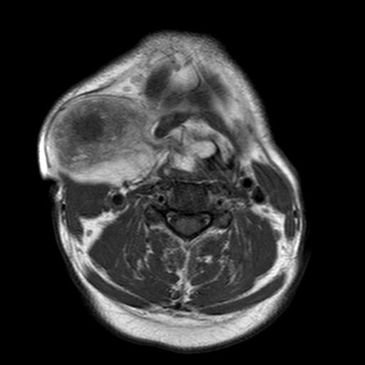
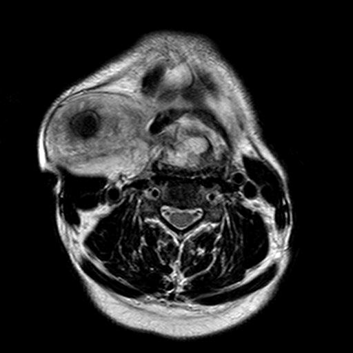
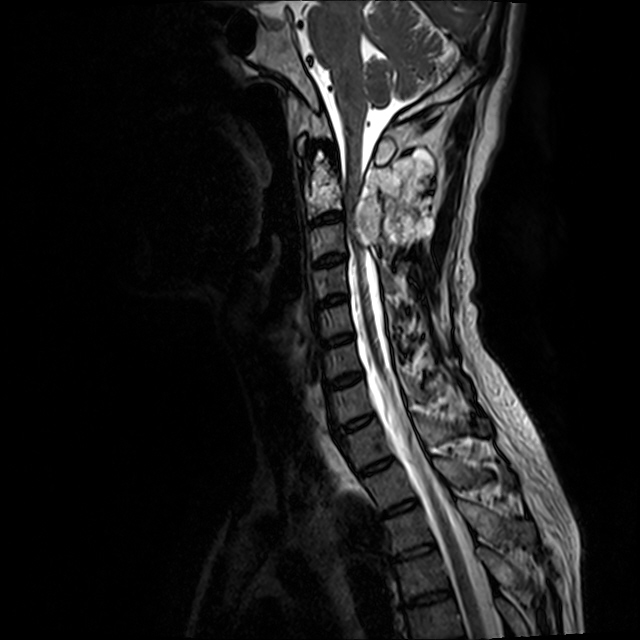
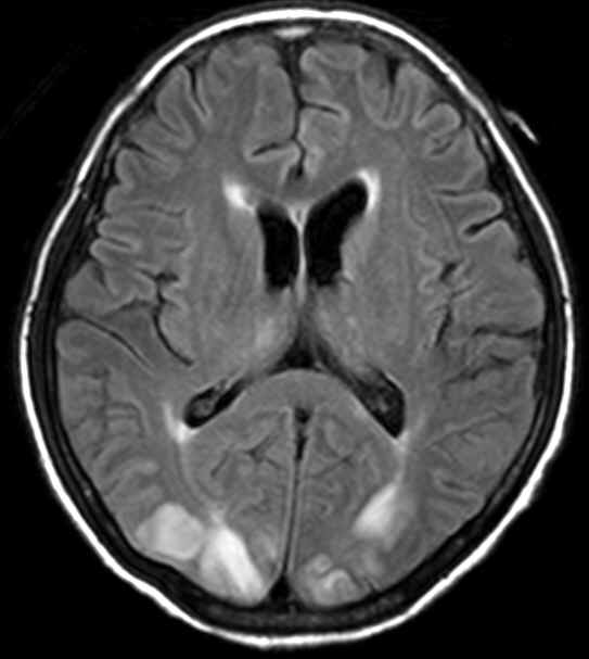
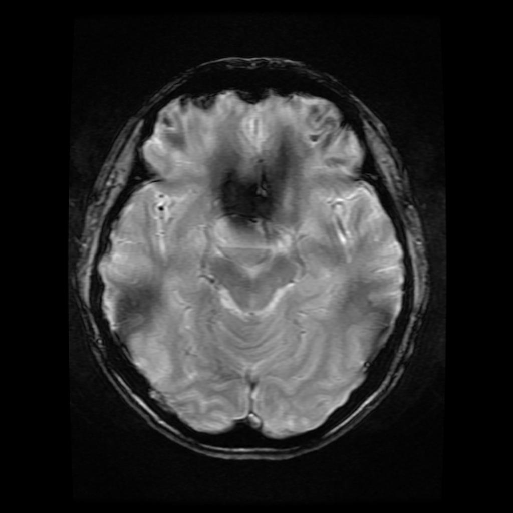
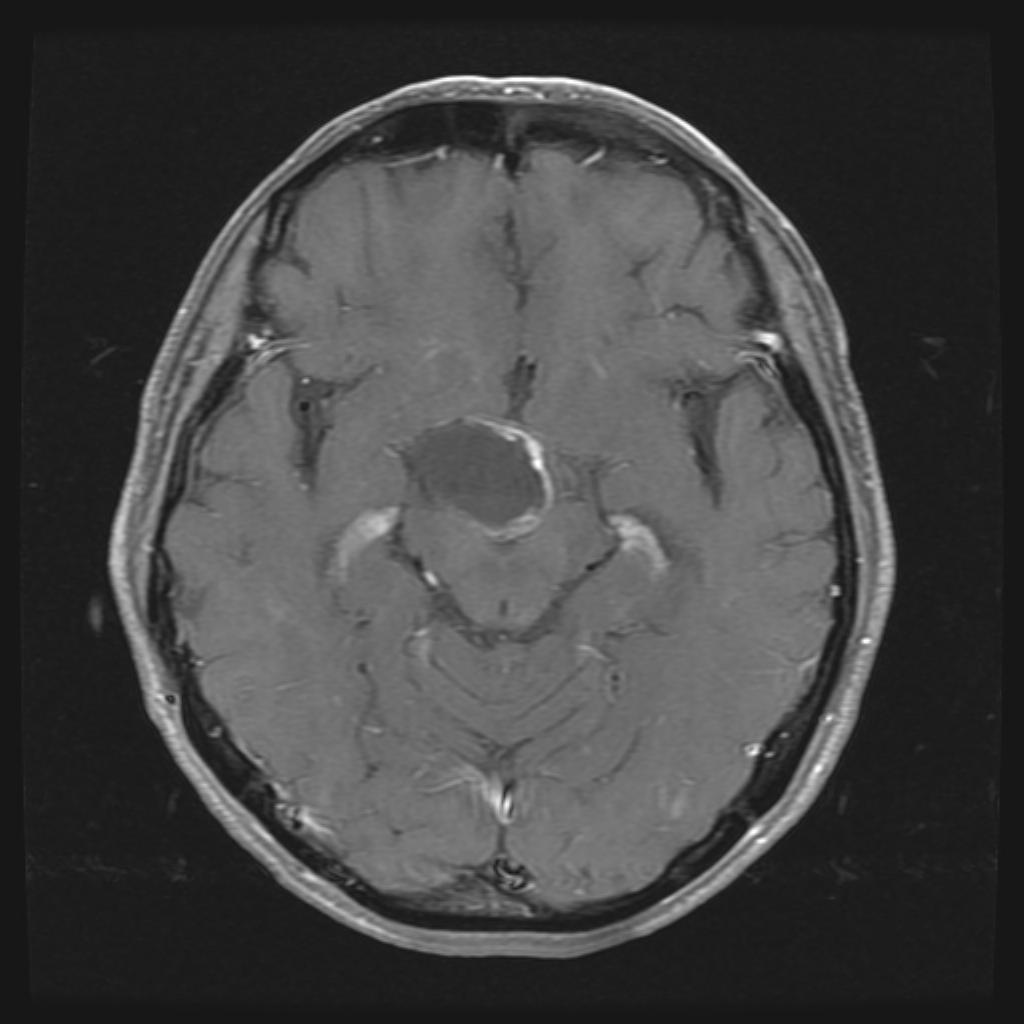
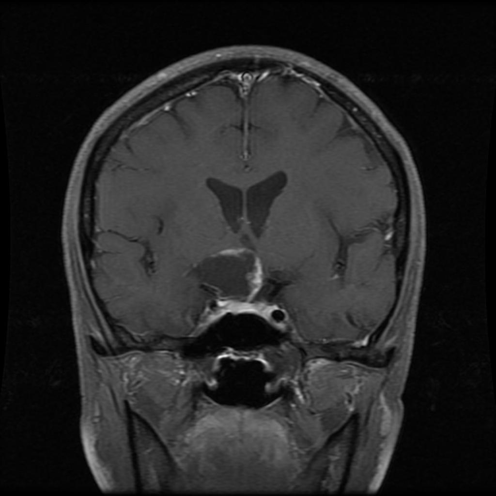
| Discussion | 진단: craniopharyngioma 영상소견:T2 sagittal image에서 주로 suprasellar area에 위치한 cystic mass가 있으며 조영증강시행하여 irregular wall enhancement를 보인다. 정상적인 뇌하수체가 보이며 mass와는 관계가 없어 보인다. Non-enhanced CT scan에서 mass의 peripheral portion에 focal calcification이 있다. Review: craniopharyngioma는 Rathke pouch squamous epithelium에서 기원한 양성종양이다. 5-15세와 50세 이후에 잘 생기는 것으로 알려져 있다. 약 70%에서 suprasellar and sellar area에 위치하고 20%에서 suprasellar area에서 그리고 나머지 10%에서 sellar area에서 위치한다. Sellar 또는 suprasellar area에 종양중 가장 많은 빈도를 차지하는 것은 뇌하수체 선종이며 뇌하주체 선종이 크기가 커져서 suprasellar extension을 하면 craniopharyngioma와 감별이 어려울 때가 있다. 하지만 CT상에서 calcification이 있을때, solid and cystic complex mass가 있을때, 조영증강 시행하여 좀 더 두꺼운 wall enhancement를 보일때 craniopharyngioma에 좀 더 합당한 소견이다. |
| Correct answer |
Correct AnswerSemi-Correct Answer |