HOME > Weekly Case > Weekly Case
Weekly Case
종결
Case No. 184
0000-00-00~0000-00-00
출제자 : 관리자
Hit : 315
| Period | 0000-00-00~0000-00-00 |
|---|---|
| Diagnosis | |
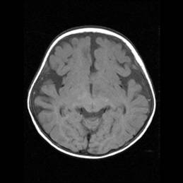
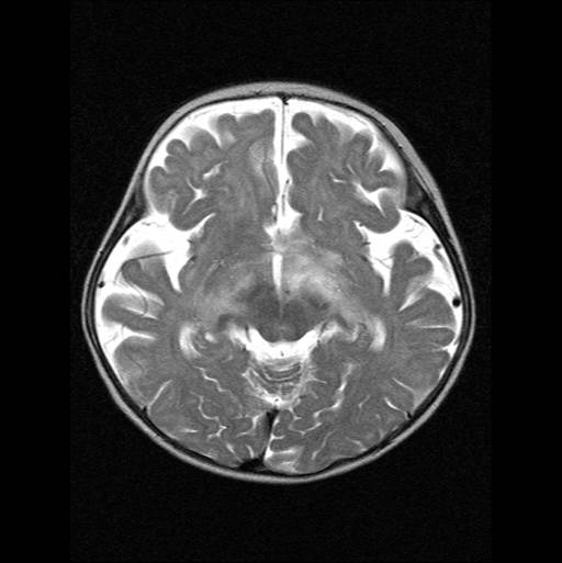
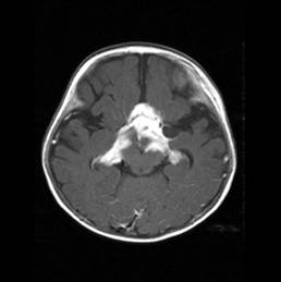
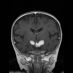
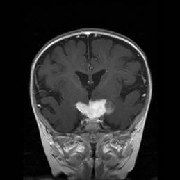
| Clinical information | 1세 여아. nystagmus로 내원. |
| Discussion | pilomyxoid astrocytoma in the optic pathway Pilomyxoid astrocytoma (PMA) is a recently described astrocytic tumor with unique histopathologic and clinical characteristics. The most pertinent features of PMA that differentiate it from typical PA are a higher rate of recurrence and CSF dissemination. PMA has a strong geographic predilection for the hypothalamic/chiasmatic region. Imaging findings in patients with PMA described a solid hypothalamic tumor with homogeneous contrast enhancement and T2 hyperintensity extending into the adjacent deep white and gray matter. A large solid hypothalamic tumor in a very young patient has been considered the classic pattern. PA and PMA are difficult to distinguish on the basis of imaging findings alone. Whereas PA is more commonly found in the posterior fossa, its second most common intracranial location is the hypothalamus/optic chiasm, the most common location of PMA. Reference 1. L.L. Linscott, A.G. Osborn et al. Pilomyxoid Astrocytoma: Expanding the Imaging Spectrum. American Journal of Neuroradiology 2008; 29:1861-1866 2. Atilla Arslanoglua, Bayram Cirak. Et al. MR Imaging Characteristics of Pilomyxoid Astrocytomas. American Journal of Neuroradiology 2003; 24:1906-1908 |
| Correct answer |
Correct AnswerSemi-Correct Answer |