HOME > Weekly Case > Weekly Case
Weekly Case
종결
Case No. 175
0000-00-00~0000-00-00
출제자 : 관리자
Hit : 276
| Period | 0000-00-00~0000-00-00 |
|---|---|
| Diagnosis | |
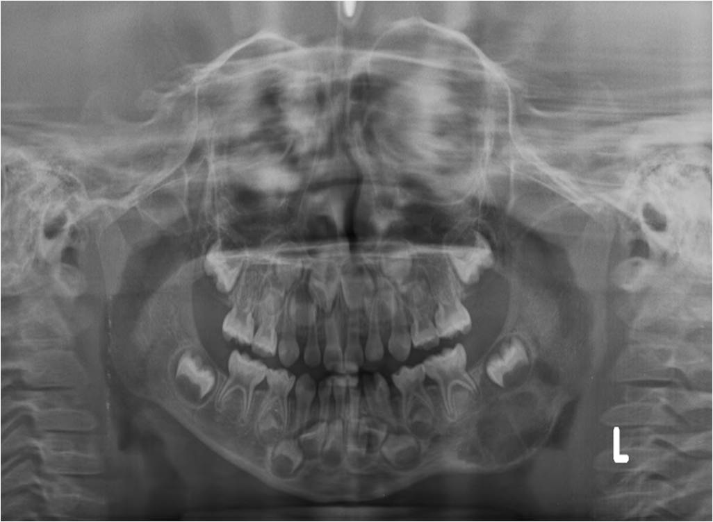
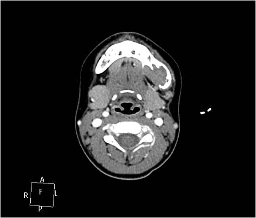
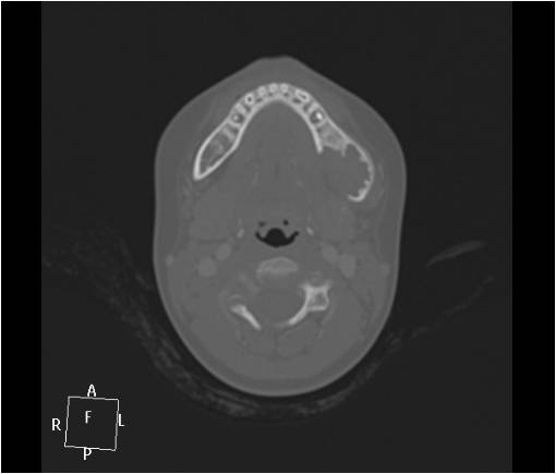
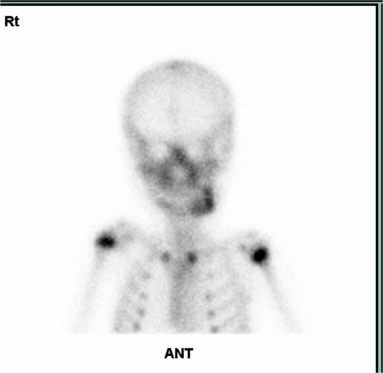
| Clinical information | 33개월 남아. 얼굴 비대칭으로 내원. |
| Discussion | Langerhans' cell histocytosis (incisional biopy of mandible) Imaging findings: About 2.5cm measured multiloculated radiolucent lesion of mandible posterior body and angle (left). Review Langerhans cell histocytosis idopathic proliferation of histiocytes leads to local tissue infiltration and destruction *3 classic syndrome 1) Eosinophilic granuloma(70%):localized benign form, isolated to bone, often monostotic 2) Hand-Schuller-Christian disease(20%) chronic form, multiple bone lesion triad : skull lesion, exophthalmos, diabetes insipidus 3) Letterer-Siwe disease(10%): acute form, multi-organ involvement, poor prognosis most often in children under 15 years of age M > F local pain, tenderness, palpable mass location: flat bone(70%) : skull(calvaria > base), mandible(body, angle),rib, pelvis, scapula, clavicle long bone(30%) : femur(m/c), humerus, tibia spine(9%) : vertebral body, thoracic > lumbar > cervical imaging appearance: well defined or poorly defined osteolytic lesion with or without sclerotic rim * Mandible: destruction of alveolar bone, erosion of the lamina dura surrounding the tooth ?밼loating tooth? Reference: N. D'Ambrosio, S. Soohoo, C. Warshall, A. Johnson, and S. Karimi Craniofacial and Intracranial Manifestations of Langerhans Cell Histiocytosis: Report of Findings in 100 Patients Am. J. Roentgenol., August 1, 2008; 191(2): 589 - 597. |
| Correct answer |
Correct AnswerSemi-Correct Answer |