HOME > Weekly Case > Weekly Case
Weekly Case
종결
Case No. 172
0000-00-00~0000-00-00
출제자 : 관리자
Hit : 323
| Period | 0000-00-00~0000-00-00 |
|---|---|
| Diagnosis | |
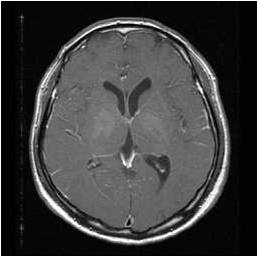
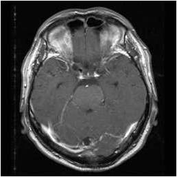
| Clinical information | 59세 남자, left side motor weakness, dysarthria로 내원 |
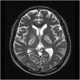
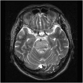
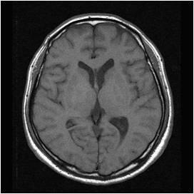
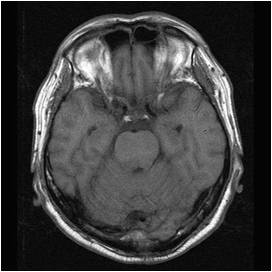
| Discussion | CNS involvement of Behcet?disease Imaging findings On T2WI, multifocal high signal intensity areas involving both periventricular white matter, posterior limbs of internal capsule, pons. They are relatively hypointense on T1WI with subtle parenchymal enhancement Review Neurobehcet disease CNS involvement of Behcet?disease Pathophysiology: small vessel vasculitis (CNS intraaxial veins; predominantly involved) Location: m/c ; brainstem, especially around the cerebral peduncles and the pons, thalamus and basal ganglia similar foci in the cerebral hemispheres MR: small foci of high signal intensity on T2WI iso- or hypointense relative on T1WI circular, linear, crescent-shaped, or irregular Non-parenchynmal involvement: Venous Thrombosis: superior sagittal sinus, transverse sinus, deep cerebral veins, cavernous sinuses Arterial Aneurysm Arterial Occlusion or Dissection Aseptic meningitis Differential diagnosis ADEM: Multifocal subcortical hyperintense foci on T2WI/ cerebral or cerebellar cortices, deep WM, BS HSV encephalitis: Limbic system (temporal lobe, insular ctx, cingulate gyri, subfrontal ctx) Behcet disease: central part of the pons/corticospinal tract, basal ganglia, thalamus, and internal capsule REFERENCE Radiologic and Clinical Findings of Beh?t Disease: Comprehensive Review of Multisystemic Involvement RadioGraphics 2008, 10.1148/r.e31 MR Features of Diseases Involving Bilateral Middle Cerebellar Peduncles, AJNR Am J Neuroradiol 24:1946?954, November/December 2003 |
| Correct answer |
Correct AnswerSemi-Correct Answer |