HOME > Weekly Case > Weekly Case
Weekly Case
종결
Case No. 170
0000-00-00~0000-00-00
출제자 : 관리자
Hit : 303
| Period | 0000-00-00~0000-00-00 |
|---|---|
| Diagnosis | |
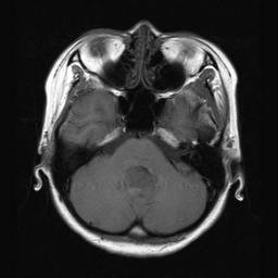
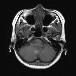
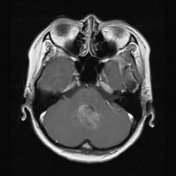
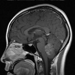
| Clinical information | 19세 여자 환자로 약 2달 전부터의 gait ataxia를 주소로 내원 |
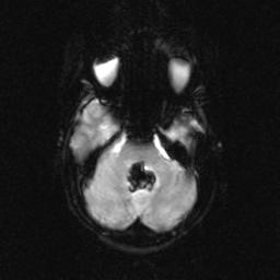
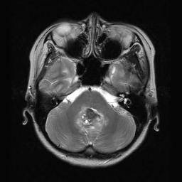
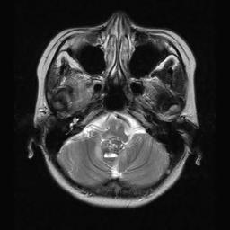
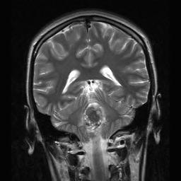
| Discussion | Medulloblastoma with confluent hemorrhage in the mass 영상소견: A heterogenous T2 dark and T1 low signal intensity mass at cerebellar vermis with compression of the 4th ventricle. After contrast enhancement heterogenous confluent enhancement is noted at the mass. On GE-EPI this mass shows fully dark signal intensity in the mass. Review: Medulloblastoma is referred to now as a primitive neuroectodermal tumor (PNET). This tumor accounts for approximately 7-8% of all intracranial tumors and 30% of pediatric brain tumors. In the brain, medulloblastoma most often arises in the posterior fossa. The tumor has the propensity of spreading throughout the CNS. Systemic metastases of this tumor, especially to bone, also have been recognized. The most common complication is hydrocephalus due to compression of the normal cerebrospinal fluid (CSF) pathways. Although this is a common complication, only 10-50% of patients with preoperative hydrocephalus will need a long-term ventricular shunt. Some children can be treated with an endoscopic third ventriculostomy. Cerebellar dysfunction: Tumor infiltration of the cerebellum usually is in the midline, leading to difficulties with ambulation and truncal ataxia. This is more common than signs attributable to the cerebellar hemisphere (eg, extremity dysmetria). Leptomeningeal dissemination: One of the most feared complications of medulloblastoma is dissemination within the CSF. Medical and, less commonly, surgical therapy must be directed at controlling dissemination to cranial nerves and spinal cord and related structures. This dissemination of disease portends to a high-risk stratification. Imaging findings: round, dense, 4th ventricle mass. As a fourth ventricle mass, it arises from roof (superior medullary velum). It is distinguishable findings from the ependymona, which arises from the floor of 4th ventricle. On NECT, it is usually hyperdense or isodense, and calcification 20%, hemorrhage rare, small intratumoral cysts/necrosis 40-50%. On CECT, it enhances very well (more than 90%) with homogenous pattern. On MR imaging, it shows T1 hypointense and T2 isointense to gray matter signals with restricted diffusion on DWI. After contrast enhancement heterogenous full enhancement is usually noted. D/Dx with teratoid-rhabdoid tumors: both atypical teratoid-rhabdoid tumors in general and infratentorial atypical teratoid-rhabdoid tumors presented at a younger age than medulloblastomas. Eleven of 19 atypical teratoid-rhabdoid tumors were infratentorial. Cerebellopontine angle (CPA) involvement was more frequent (8/11, 72.7%) in atypical teratoid-rhabdoid tumor than in medulloblastoma (4/36, 11.1%) (p < 0.001). Intratumoral hemorrhage was more common in atypical teratoid-rhabdoid tumor (9/19, 47.4%) than in medulloblastoma (2/36, 5.6%) (p < 0.0001). All atypical teratoid-rhabdoid tumors and all medulloblastomas for which DWI was available displayed increased signal intensity on DWI compared with normal brain parenchyma. The mean ADC values for tumor types were not significantly different. CONCLUSION: Atypical teratoid-rhabdoid tumor presents at a younger age than medulloblastoma. -- Koral K et al. Imaging characteristics of atypical teratoid-rhabdoid tumor in children compared with medulloblastoma. AJR Am J Roentgenol. 2008 Mar;190(3):809-14. |
| Correct answer |
Correct AnswerSemi-Correct Answer |