HOME > Weekly Case > Weekly Case
Weekly Case
종결
Case No. 169
0000-00-00~0000-00-00
출제자 : 관리자
Hit : 284
| Period | 0000-00-00~0000-00-00 |
|---|---|
| Diagnosis | |
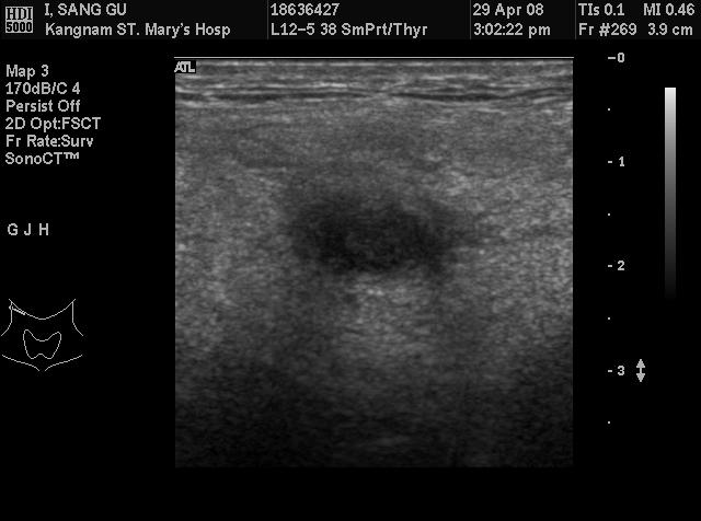
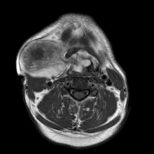
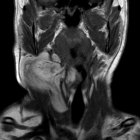
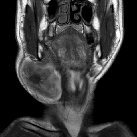
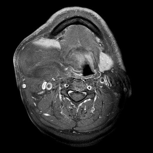
| Clinical information | 50세 남자환자로 수년 전부터 만져지는 우측 경부의 종괴를 주소로 내원 |
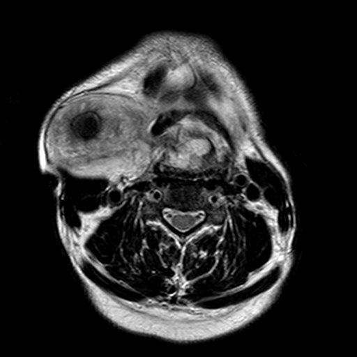
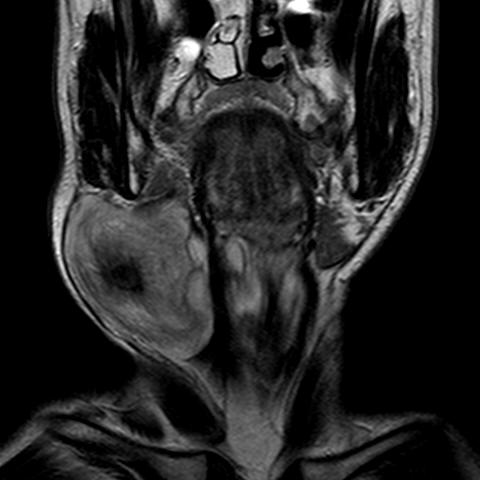
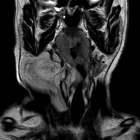
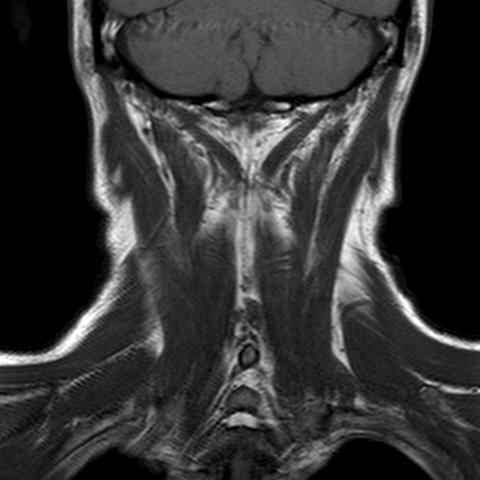
| Discussion | Spindle cell lipoma 영상소견: A large and diffuse T1 high signal intensity mass involving R neck with encroachment to larynx. In the central portion of the mss, focal nodular T1 low signal intensity is noted with peri-nodular low signals. After contrast enhancement focal enhancement is noted at this T1 low nodular lesion. Review: Spindle cell lipoma was first described by Enzinger and Harvey in 1975 as a benign lesion in which mature fat is replaced by collagen-forming spindle cells. Knowledge of this appearance is important because as a result of the variable ratio of adipose and nonadipose soft tissue in these tumors, a spindle cell lipoma may mimic a liposarcoma radiologically. In a 10-year review of soft-tissue tumors seen at the Armed Forces Institute of Pathology, spindle cell lipoma was encountered only slightly less frequently than well-differentiated liposarcoma. Spindle cell lipoma is a benign lesion that is cured by local excision and has never been reported to metastasize. These slow-growing, usually solitary, painless lipomatous variants typically present in men between the ages of 45 and 65 years. In their original report, Enzinger and Harvey noted that these lesions typically occur in men, accounting for 91% (104/114) of their patients. This trend was substantiated in a subsequent Armed Forces Institute of Pathology report of 816 patients, in which 88% were men. In keeping with these reports, all the patients in our study were men. Spindle cell lipoma also has a significant tendency to occur in the subcutaneous tissue of the posterior neck, shoulder, and back. In the two large series previously noted, the posterior neck and head and neck accounted for 36% and 41% of cases, respectively. Tumors have also been reported in the extremities, oral cavity, larynx, bronchus, orbit, scalp, breast, and perianal region. Before its designation as a separate entity, spindle cell lipoma was frequently misdiagnosed pathologically as liposarcoma. Microscopically, the lesion is composed of mature fat with areas replaced by fibroblast-like spindle cells within a matrix of mucin and collagen fibrils. Although most spindle cell lipomas are composed of a relatively equal ratio of fat and spindle cells, either component may predominate, and the variation in the ratio of fat and spindle cells causes the wide spectrum of imaging features. Prominent vascularity most likely accounts for the intense enhancement in the nonadipose components of the spindle cell lipomas. This enhancement is significantly more intense than that seen in liposarcoma. The imaging appearance of spindle cell lipoma is not pathognomonic and, like most other musculoskeletal tumors, displays a spectrum of features. In the case of spindle cell lipoma, these features may overlap with those of liposarcoma. The diagnosis of spindle cell lipoma, however, should be suggested when a middle-aged man presents with a well-defined complex fatty mass in the subcutis, especially when localized to the posterior neck. Intense enhancement of the nonadipose component further supports this diagnosis. Bancroft LW et al. Imaging characteristics of spindle cell lipoma. AJR Am J Roentgenol. 2003 Nov;181(5):1251-4. |
| Correct answer |
Correct AnswerSemi-Correct Answer |