HOME > Weekly Case > Weekly Case
Weekly Case
종결
Case No. 168
0000-00-00~0000-00-00
출제자 : 관리자
Hit : 286
| Period | 0000-00-00~0000-00-00 |
|---|---|
| Diagnosis | |
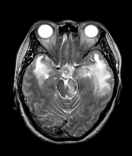
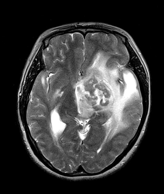
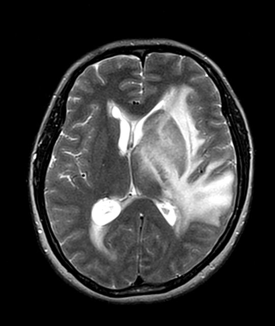
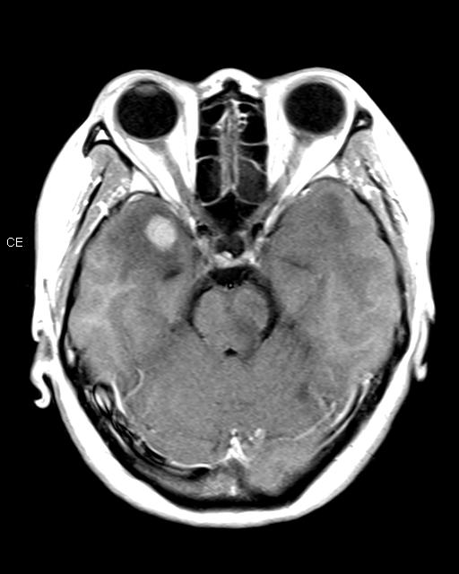
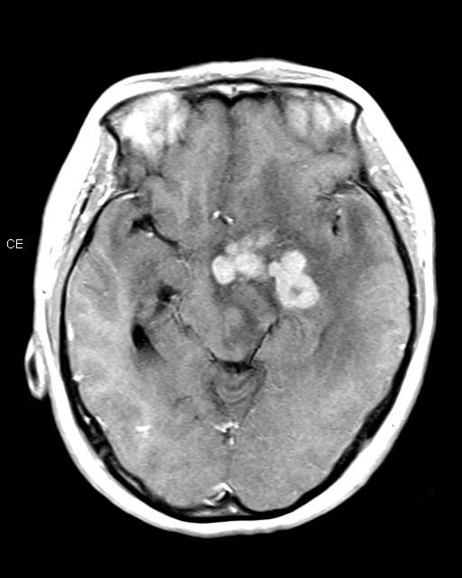
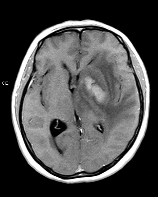
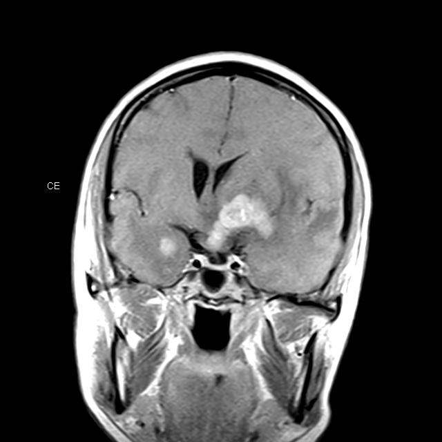
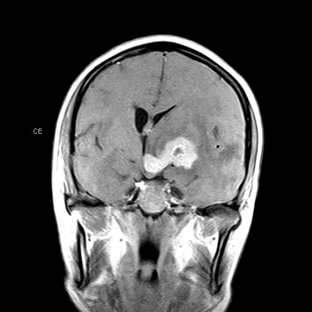
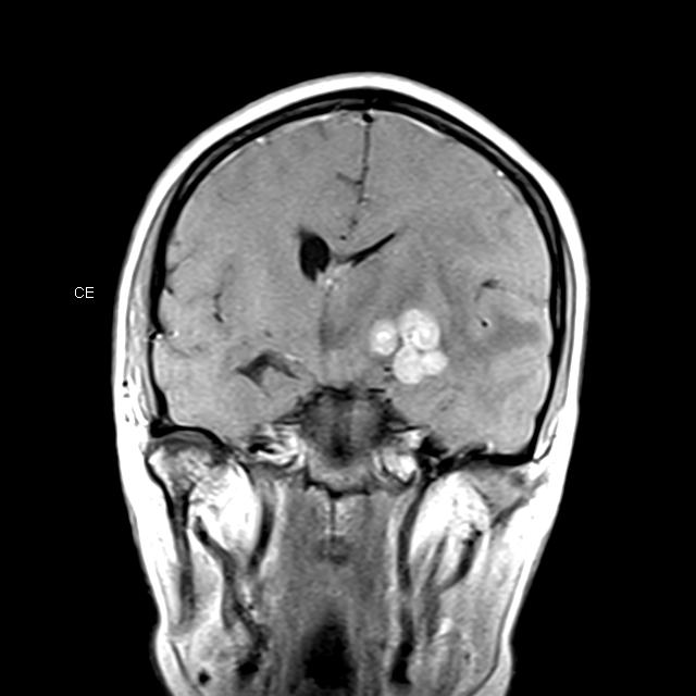
| Clinical information | 49세 여자환자로 약 한 달 전부터 headache, insomnia, intermittent disorientation 있어 정신과 내원. 내원 후 falling tendency 발생하여 brain MRI 시행 |
| Discussion | CNS Lymphoma (large B cell type) 영상소견: A large multi-nodular enhancing masses involving L medial temporal lobe, basal ganglia, hypothalamus, 3rd ventricle lateral wall with confluent edema on adjacent white matter. And another enhancing nodular mass at anterior portion of R temporal lobe. Review: Roman-Goldstein SM et al. MR of primary CNS lymphoma in immunologically normal patients AJNR Am J Neuroradiol. 1992 Jul-Aug;13(4):1207-13. Primary CNS lymphoma presented as solitary enhancing lesions in 40% of the patients and multiple lesions in 40%. Thirty-three separate lesions were visible: 58% abutted the ventricular system, 76% showed a homogenous enhancement pattern, and 79% showed marked enhancement. Primary CNS lymphoma usually presents as solitary or multiple dense homogenous enhancing lesions that abut an ependymal surface. These lesions can be divided into an enhancing area and an area of surrounding abnormal signal. These two areas often have different signal intensities on unenhanced T2-weighted images. These findings are sufficiently suggestive of the diagnosis of primary CNS lymphoma that a needle biopsy be performed based on these findings. Coulon A et al. Radiographic findings in 37 cases of primary CNS lymphoma in immunocompetent patients. Eur Radiol. 2002 Feb;12(2):329-40. Epub 2001 Sep 21. Primary central nervous system lymphoma presents as supratentorial solitary lesions in approximately 80% of the patients and multiple lesions in 20%. In contrast to classical data, the lesions are located in deep structures only in one-third of the cases, and involve posterior fossa in 10% of cases. Most of the lesions are hyperdense or isodense (92%) on CT, hypointense or isointense on T1-weighted images, and only about 40% are hyperintense on T2-weighted images. Nearly all the lesions enhance, except after corticosteroid administration. They produce mild oedema and mass effect. Meningeal or ventricular enhancement are rare but suggestive. Calcification, haemorrhage or necrosis are scarce. Although PCNSL in immunocompetent patients have a variable CT and MR appearance, the imaging data often suggest the diagnosis |
| Correct answer |
Correct AnswerSemi-Correct Answer |