HOME > Weekly Case > Weekly Case
Weekly Case
종결
Case No. 164
0000-00-00~0000-00-00
출제자 : 관리자
Hit : 344
| Period | 0000-00-00~0000-00-00 |
|---|---|
| Diagnosis | |
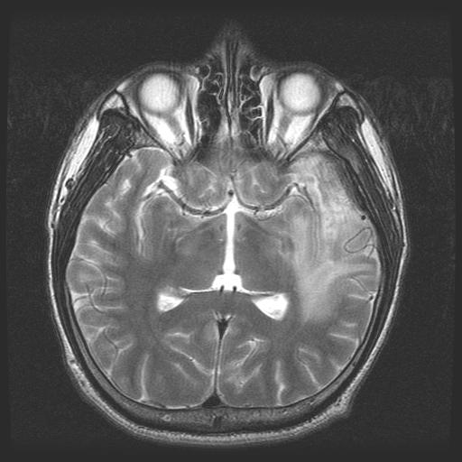
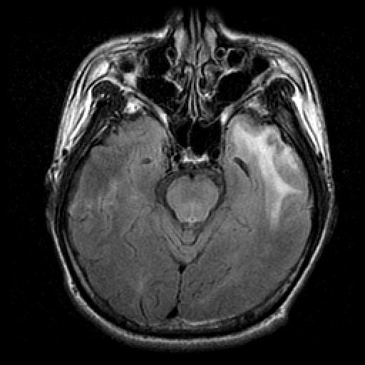
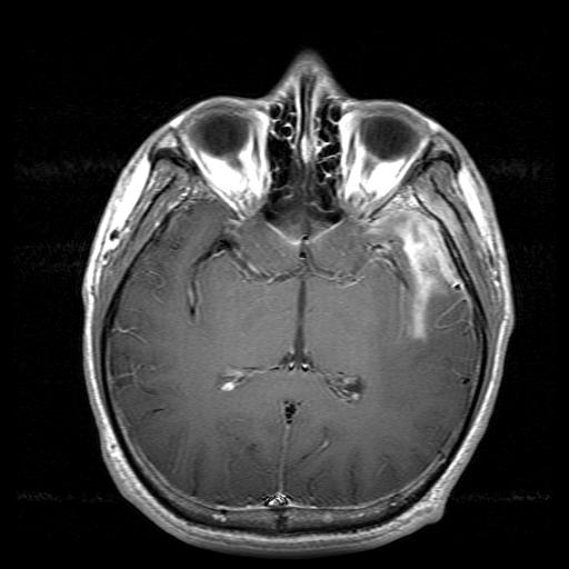
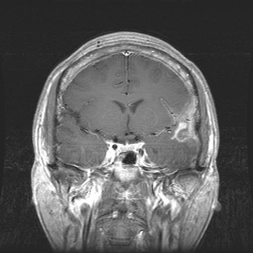
| Clinical information | 29세 남자, aphasia, seizure로 검사 시행 |
| Discussion | Meningeal inflammatory myofibroblastic tumor (inflammatory pseudotumor) 좌측 frontotemporal convexity에 pachymeningeal thickening과 enhancement 및 인접한 leptomeninges의 enhancement 소견이 있으며, T2WI에서 pachymeningeal lesion은 전반적으로 dark SI를 보인다. 동시에 인접한 sphenoid bone의 greater wing과 temporalis muscle의 enhancement 소견도 있다. Inflammatory myofibroblastic tumor는 병리적으로 모호한 disease entity이며, 특히 CNS에서 이전에는 inflammatory pseudotumor 혹은 plasma cell granuloma로 주로 불리어 왔으나, 최근에 병리적으로 inflammatory myofibroblastic tumor로 정리되고 있는 추세이다. CNS inflammatory myofibroblastic tumor는 드물지만, 곳곳에서 발생하는 것으로 보고되었고 이중 meningeal origin이 상대적으로 흔한 것으로 되어 있다. 병리적인 definition이 pseudotumor에서 tumor 쪽으로 정리되고 있다는 점이 시사하듯이 tumor의 aggressive feature를 보일 수 있으며, recurrence도 생길 수 있다. 주로 pachymenigeal origin이지만, 인접 leptomeningeal involvement도 동반될 수 있으며, dural sinus thrombosis 및 mastoid involvement 등도 가능하다. 병변의 T2 low SI가 비교적 특징적이라 할 수 있지만, localized or diffuse dural thickening으로서 T2 low SI를 보일 수 있는 lymphoma, fibroblastic meningioma를 포함하여, idiopathic hypertrophic pachymeningitis와 여러 원인의 infectious meningitis 등이 유사한 feature를 보일 수 있다. Han MH, Chi JG, Kim MS, et al. Fibrosing inflammatory pseudotumors involving the skull base: MR and CT manifestations with histopathologic comparison. AJNR Am J Neuroradiol 1996; 17:515-521. |
| Correct answer |
Correct AnswerSemi-Correct Answer |