HOME > Weekly Case > Weekly Case
Weekly Case
종결
Case No. 163
0000-00-00~0000-00-00
출제자 : 관리자
Hit : 317
| Period | 0000-00-00~0000-00-00 |
|---|---|
| Diagnosis | |
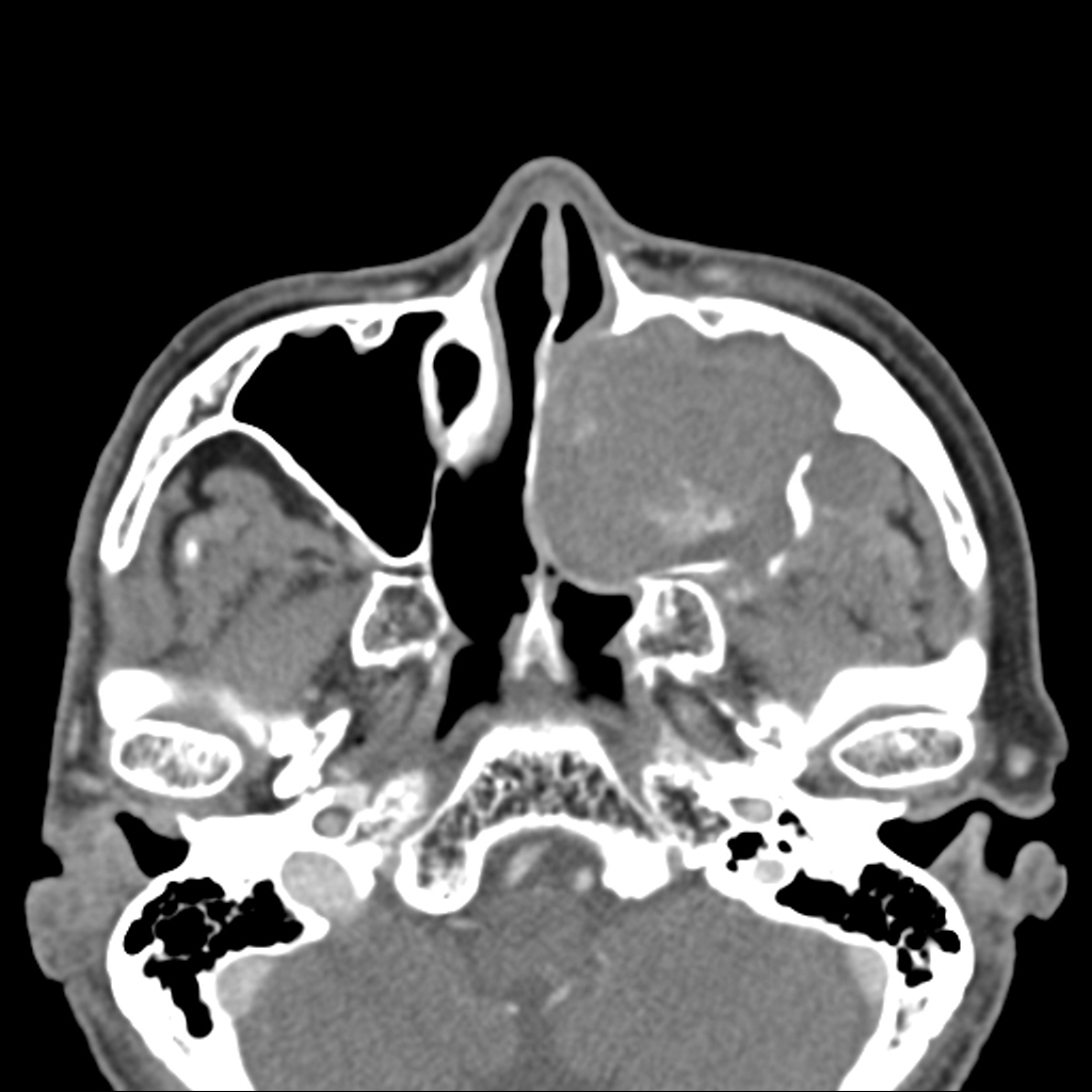
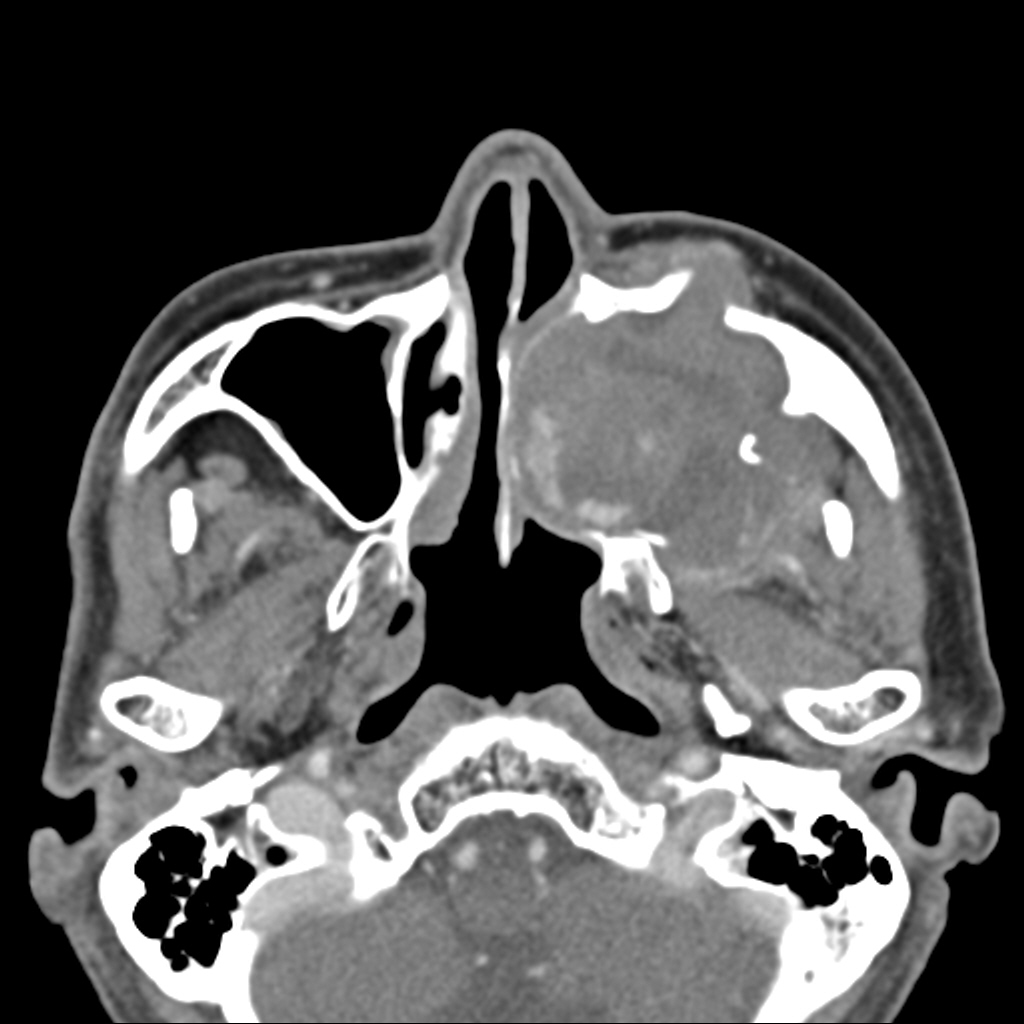
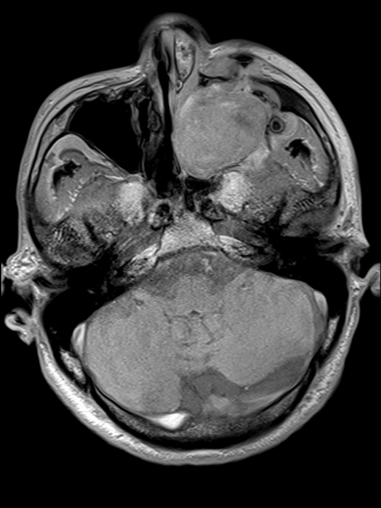
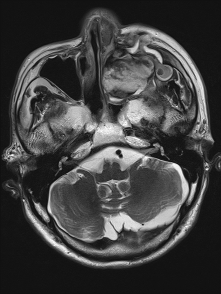
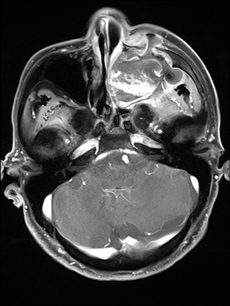
| Clinical information | 86세 남자. 1개월 전부터 시작된 cheek swelling 발생하였고, left inferior meatus bulging mass로 암 의심하에 내원함. |
| Discussion | Organizing hematoma of left maxillary sinus pathophysiology: Trauma, surgery, bleeding diasteheses등으로 인한 maxillary sinus내의 혈괴 축적 poor ventilation & drainage fibrous capsule형성, 흡수가 방해되고, neovascularization, 재출혈, fibrosis imaging appearance: sinonasal OH는 expansile soft tissue mass with erosion of sinus wall로서 T2 hypointense rim을 가지고, patchy, papillary, or frondlike enhancement를 가진다. DDx. mucocele, fungus ball, inflammatory polyp, cholesterol granuloma, inverted papilloma, sinonasal hemangioma References: Sinonasal Organized Hematoma: CT and MR imaging Findings AJNR AJNR Am J Neuroradiol 2006;29:1204-1208 Expansile Organized Maxillary Sinus Hematoma: MR and CT Findings and Review of Literature AJNR Am J Neuroradiol 2007; 28:1375–1377 Organized Hematoma of the Maxillary Sinus: CT Findings AJR 2007; 188:W370–W373 |
| Correct answer |
Correct AnswerSemi-Correct Answer |