HOME > Weekly Case > Weekly Case
Weekly Case
종결
Case No. 161
0000-00-00~0000-00-00
출제자 : 관리자
Hit : 301
| Period | 0000-00-00~0000-00-00 |
|---|---|
| Diagnosis | |
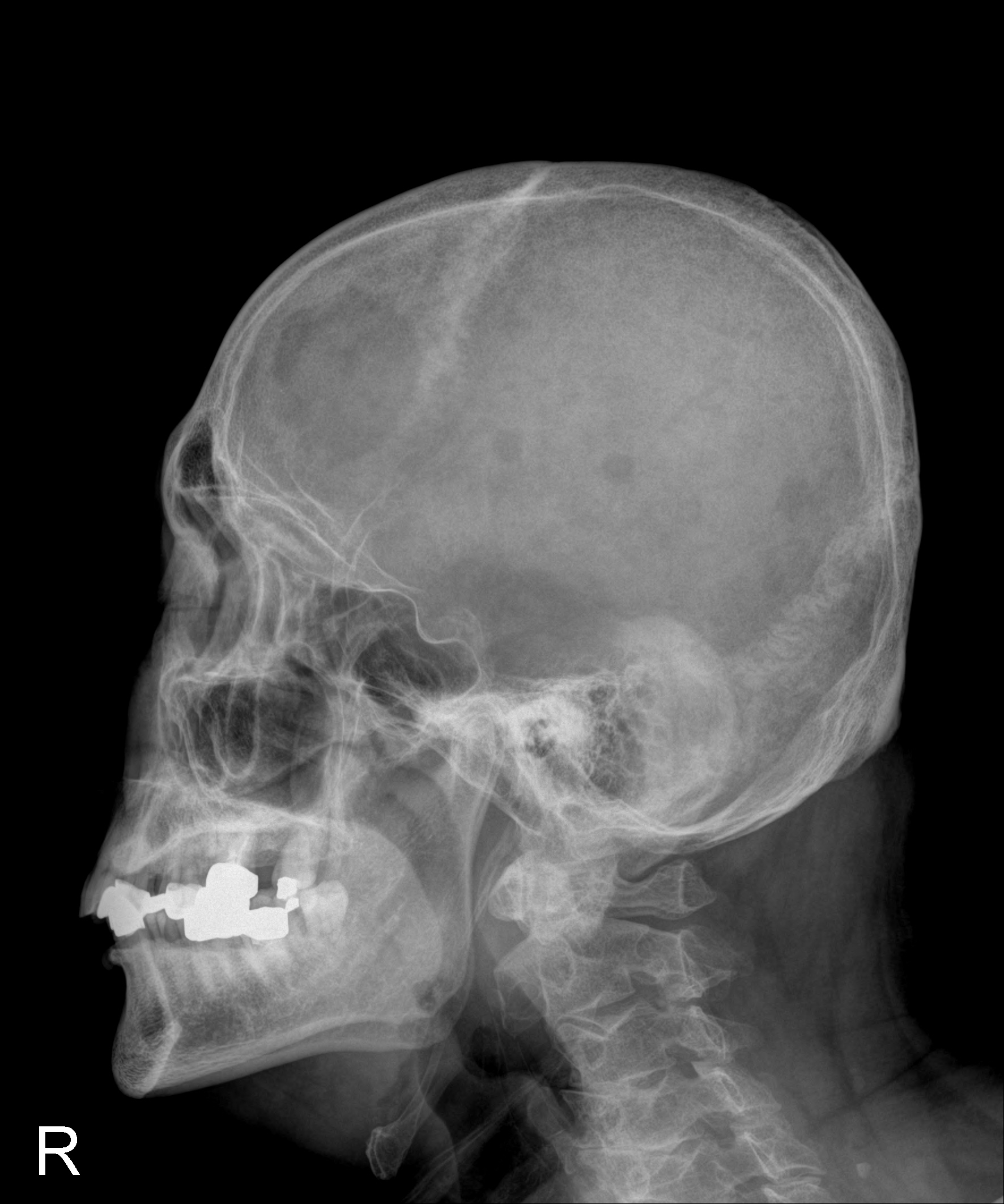
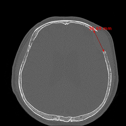
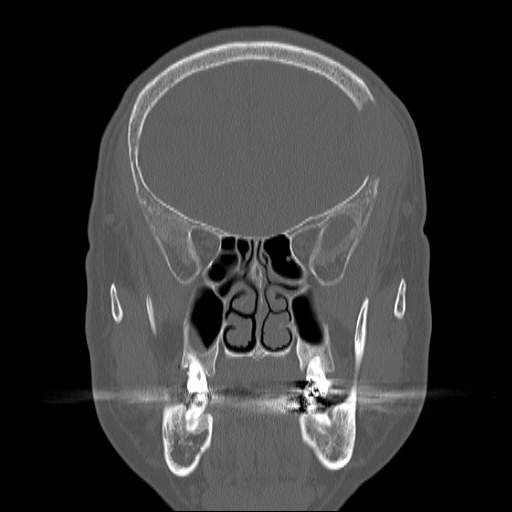
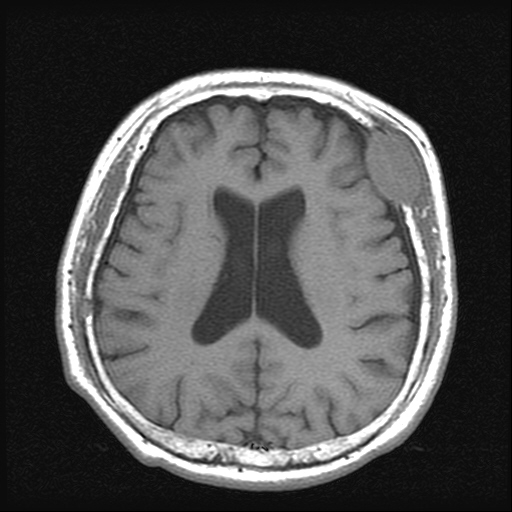
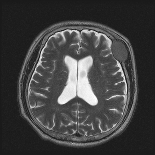
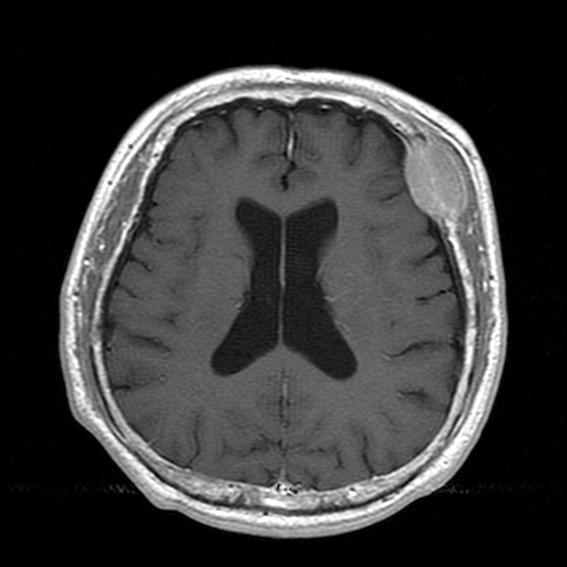
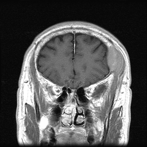
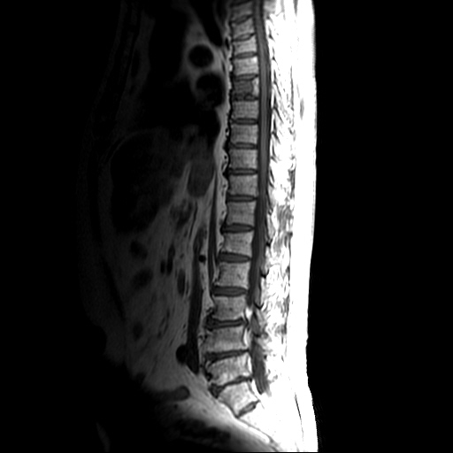
| Clinical information | 69세 남자. Right femur neck fracture에 대한 evaluation도중 발견한 scalp mass. |
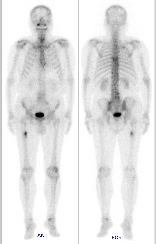
| Discussion | Multiple myeloma involvement of T-spine & skull (bone marrow biopsy및 skull biopsy에 의하여 모두 confirmed됨) Definition: multifocal malignant proliferation of monoclonal plasma cells within bone marrow Imaging findings: Multifocal, diffuse, or heterogeneous T1hypointensity Location: axial skeleton (red marrow)> long bones Spine (87% vertebral compression Fx between T6 and L4), skull, ribs, pelvis Size: Variable Well-circumscribed, punched-out lesions on plain-radiograph Radiographic findings Plain: Diffuse osteropenia (85%), multiple osteolytic lesions (80%) CT: Multifocal osteolytic lesions/ vertebral compression Fx or destruction, extraosseous soft tissue component MR: Normal Bone marrow involvement with focal, diffuse or variegated patterns Nuclear Medicine Bone scan: typically negative, only 10% detected on bone scan PET: metabolically active Sensitivity: 84-92%, specificity: 83-100% Reference: Diagnostic imaging Spine Amirsys press IV:1-66 to 68 |
| Correct answer |
Correct AnswerSemi-Correct Answer |