HOME > Weekly Case > Weekly Case
Weekly Case
종결
Case No. 151
0000-00-00~0000-00-00
출제자 : 관리자
Hit : 338
| Period | 0000-00-00~0000-00-00 |
|---|---|
| Diagnosis | |
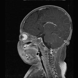
| Clinical information | 생후 2개월 신생아 (남자). 양측 눈동자가 밝게 보여 내원. 미숙아 등 특이 과거력 없음. |
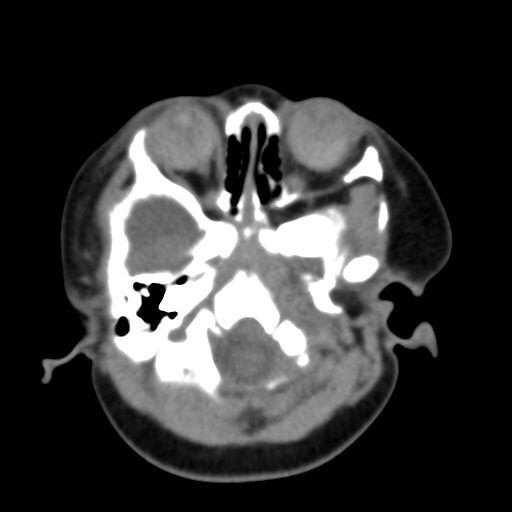
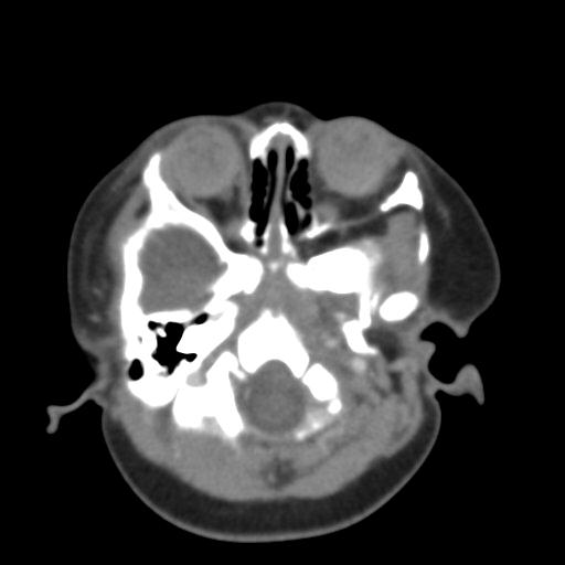
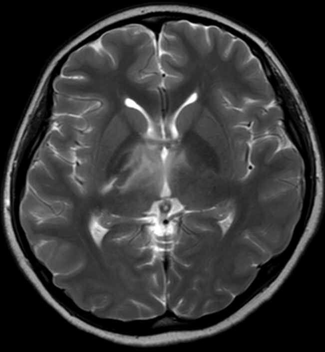
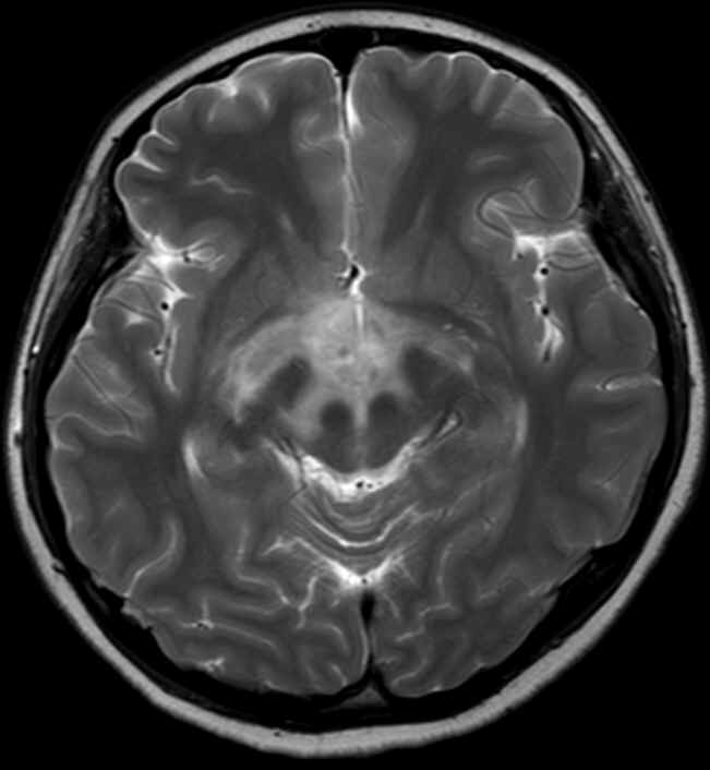
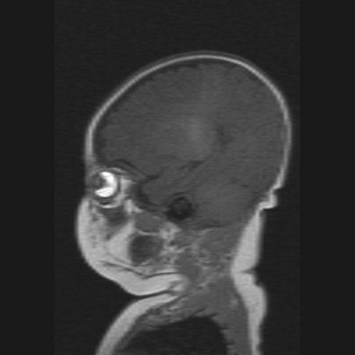
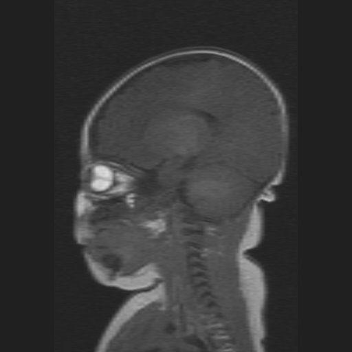
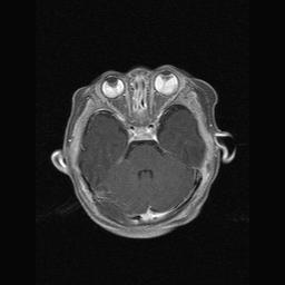
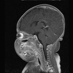
| Discussion | Persistent hyperplastic primary vitreous * 영상소견 조영증강전 CT영상에서 양쪽 안구내 음영이 전반적으로 증가되어 있고 calcification은 동반되어 있지 않으며 조영증강되는 종괴는 없음. MR영상에서 retinal detachment가 있고 T2WI low, T1WI high signal을 보여 subretinal hemorrhage가 의심되며 orbital cavity내에 명확한 종괴형성이 없으며 lens에서 retina까지 연결되는 stalk like lesion이 있음. * Review 우측 안구를 total vitrectomy시행하였으며 total retinal detachment와 subretinal fluid and hemorrhage가 있었고 proliferative membranous structure와 stalk이 발견되었다. Leukokoria의 원인에는 retinoblastoma, PHPV, coat disease, retinopathy of prematurity(ROP), toxocaral endophthalmitis가 있다. PHPV는 embryonic ocular blood supply를 담당하는 primary vitreous(embryonic hyaloid vasculature)의 incomplete regression에 의한 congenital ocular lesion이다. 대개는 unilateral involvement를 하며 small globe를 보이는 것이 특징고 retinal detachment를 흔히 동반한다. CT상 hyperdense하게 보이고 calcification이 없다. MR상 vitreous가 T1WI & T2WI에서 고신호강도로 보이며 다양한 blood product에 의한 signal이 다양하게 보일 수 있다. Coat disease는 almost always unilateral lesion으로 알려져 있으며 ROP의 경우 premature infant에서 supplemental oxygen therapy의 기왕력이 있어야 한다. Radiographics 1994;14:1059-1079 |
| Correct answer |
Correct AnswerSemi-Correct Answer |