HOME > Weekly Case > Weekly Case
Weekly Case
종결
Case No. 147
0000-00-00~0000-00-00
출제자 : 관리자
Hit : 306
| Period | 0000-00-00~0000-00-00 |
|---|---|
| Diagnosis | |
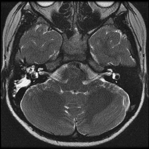
| Clinical information | 11세 여아. 우측 청력 감소로 내원. |
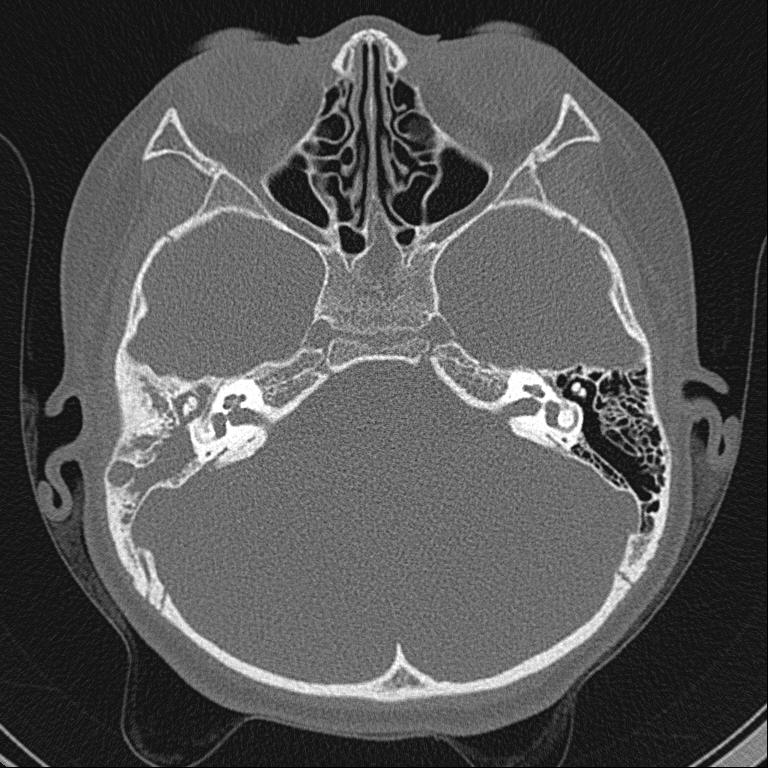
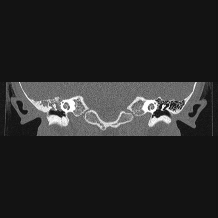
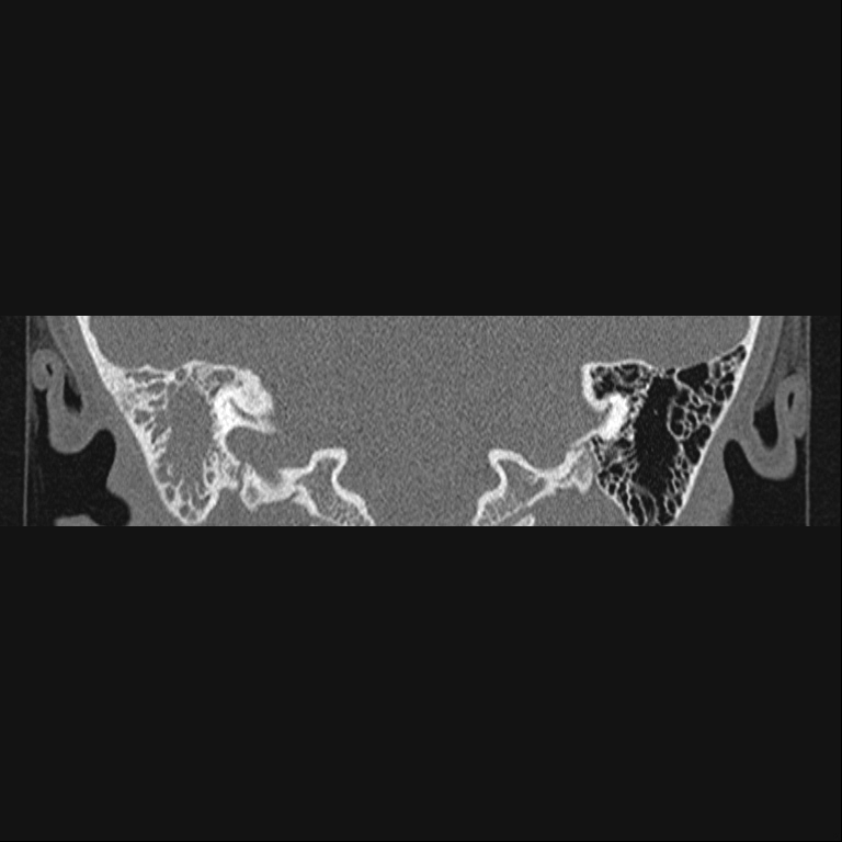
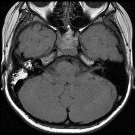
| Discussion | Cholesterol granuloma, middle ear Imaging findings: Smooth mass of high T1 & T2 signal in mastoid antrum and middle ear Reference: Cholesterol granuloma, middle ear ?Recurrent hemorrhage into middle ear cavity cause inflammatory mass of granulation tissue ?Slowly progressive conductive hearing loss ?Otoscopy: non-pulsating bluish discoloration of tympaninc membrane = blue ear drum ?DDx: dehiscent jugular bulb, aberrant internal carotid artery, COM with hemorrhage ?Occur throughout body, including petrous apex & orbit ?More commonly arise in middle ear NECT ?Early findings: small middle ear mass, no ossicular loss ?Late findings ?Opacified middle ear & mastoid, ossicular diplacement ?Expansible bony change with scalloping of surrounding bone ?CECT: DDx form glomus tympanicum paraganglioma ?MR ?T1WI: high signal (paramagnetic effect of methemoglobin) ?T2WI: central high signal from granulation tissue and peripheral low signal from hemosiderin deposit ?MRA: useful to distinguish from vascular anomalies N Martin, O Sterkers, D Mompoint, N Julien, and H Nahum Cholesterol granulomas of the middle ear cavities: MR imaging Radiology 1994; 191: 249. |
| Correct answer |
Correct AnswerSemi-Correct Answer |