HOME > Weekly Case > Weekly Case
Weekly Case
종결
Case No. 145
0000-00-00~0000-00-00
출제자 : 관리자
Hit : 289
| Period | 0000-00-00~0000-00-00 |
|---|---|
| Diagnosis | |
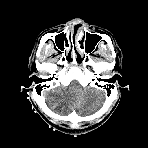
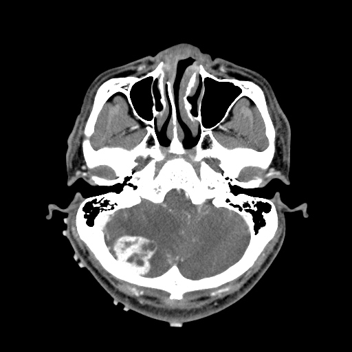
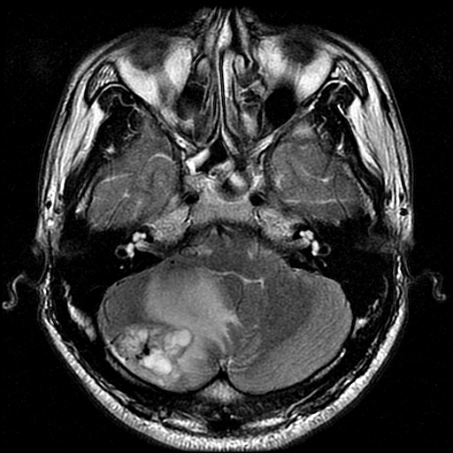
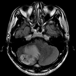
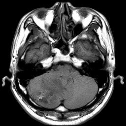
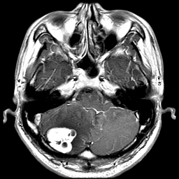
| Clinical information | 31세 남자. 1개월간의 두통과 어지러움으로 내원. 과거력상 pheochromocytoma 진단 받음. |
| Discussion | Hemangioblastoma Imaging findings: ?Intra axial posterior fossa mass ?Some cystic lesions with well enhancement ?Highly vascularized mass on DSA Reference: Hemangioblastoma ?Incidence ?7% of 1?posterior fossa tumor in adult ?Sporadic or VHL (10~20%) ?Age ?3rd~5th decade, rare in children ?Location ?80~85%; Cbll ?3~13%; spinal cord ?Supratentorial; uncommon ?Curable by surgery von Hippel-Lindau Syndrome ?Autosomal dominant ?Variety of benign & malignant tumors ?Hemangioblastoma including retinal angioma; 60~80% ?RCC; m/s cause of death ?Pheochromocytoma ?Endolymphatic sac tumor of middle ear ?Serous cystadenoma & neuroendocrine tumor of pancreas ?Papillary cystadenoma of epididymis ?VHL type I ?No pheochromocytoma ?VHL type II ?High risk of pheochromocytoma ?Type IIA; low risk of RCC ?Type IIB; high risk of RCC ?Type IIC; only pheochromocytoma without hemangioblastoma & RCC |
| Correct answer |
Correct AnswerSemi-Correct Answer |