HOME > Weekly Case > Weekly Case
Weekly Case
종결
Case No. 144
0000-00-00~0000-00-00
출제자 : 관리자
Hit : 328
| Period | 0000-00-00~0000-00-00 |
|---|---|
| Diagnosis | |
| Clinical information | 28세 여자. 8 개월간 지속된 몸 전체의 tremor로 내원. |
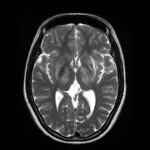
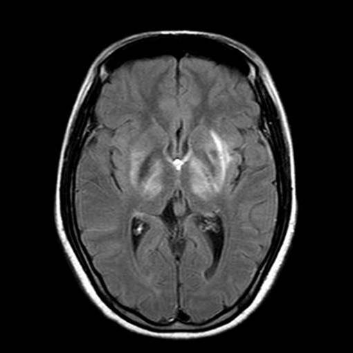
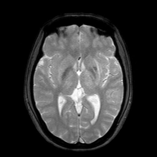
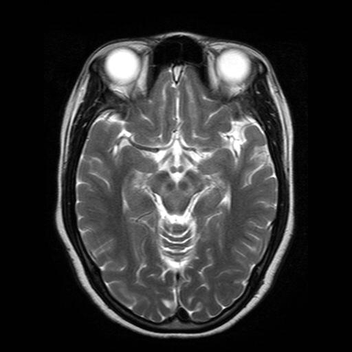
| Discussion | Wilson disease 영상소견 T2강조영상, FLAIR, T2* GRE에서 대칭적인 high signal intensity lesion이 basal ganglia, pons, thalamus, mid brain에 관찰되며 Lt putam에는 focal low signal intensity portion이 관찰됨. Review 원인: 체내 구리대사의 선천적이상이며 구리의 운반단백질인 ceruloplasmin 결핍에 의한 구리의 침착 Location: Liver, Lentiform nucleus Age: 진단시 환자의 나이는 평균 18세로 2nd -3rd decade 영상소견 MR: T2 강조영상에서 기저핵 (특히 피각과 미상핵), 시상, 뇌교, 중뇌에 양측으로 대칭성의 고신호 강도. 양측 기저핵이나 시상에 저신호 강도를 보일 수 있는데 이는 구리 침착 자체나 철분등의 상자성 물질에 의한것으로 알려짐. “Face of giant panda” sign”(중뇌병변) 확산강조영상에서 neurologic symptom이 있은 직후 시행하면 양측 basal ganglia에 고신호 강도을 보이며 추적관찰에서 low signal intensity를 보임. 참고문헌 1. Diagnostic imaging, Brain, Osborn.I:9-70~73 2. 신경방사선학. 장기현. 473-474 3. Cranial MR in Wilson Disease: Abnormal White Matter in Extrapyramidal and Pyramidal Tracts. Am J Neuroradiol 1995;16:2021–2027 4. Diffusion MR Imaging Changes Associated with Wilson Disease. Am J Neuroradiol 2003;24:965– 967 5. Wilson Disease: Findings at MR Imaging and CT of the Brain with Clinical Correlation. Radiology 1996; 198:531-536 |
| Correct answer |
Correct AnswerSemi-Correct Answer |